


We present the case of a 55-year-old woman who came to the clinic due to neck and lower back pain for the past 6 months. Physical examination of the locomotor system was normal.
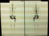
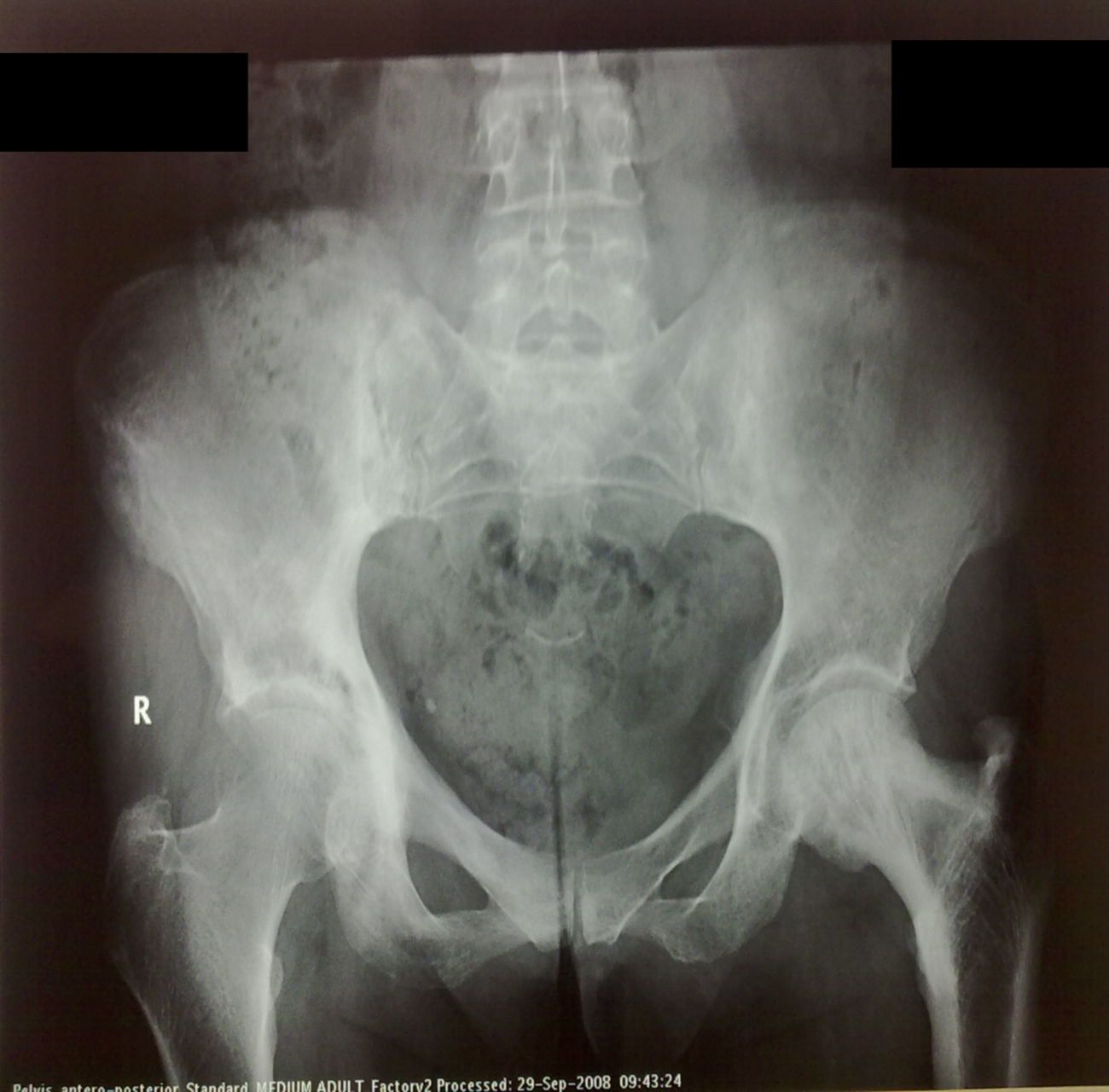
A cervical spine X-ray was taken and showed a straightening of its physiologic lordosis. Lumbar spine X-ray showed a sclerotic pattern on sacroiliac joints (Fig. 1).
Laboratory tests showed an increase in alkaline phosphatase in the blood chemistry (485UI/l) and normal serum calcium.
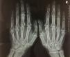
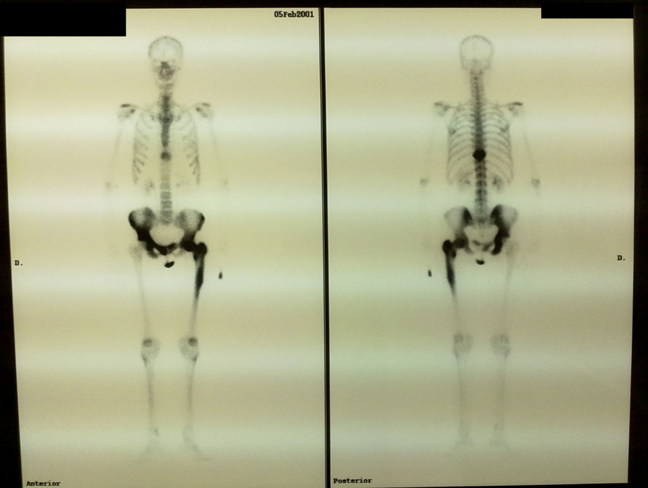
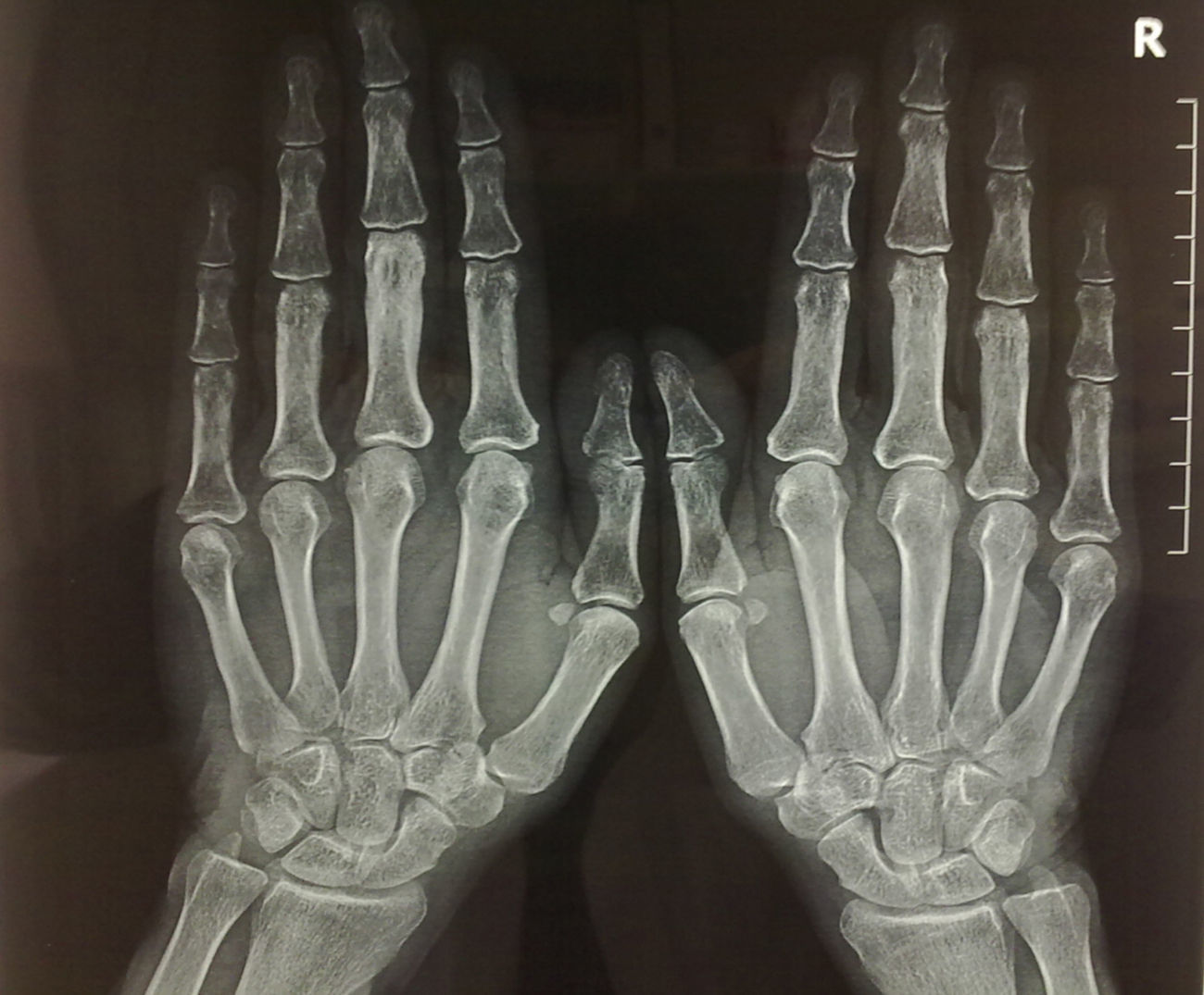
Suspecting Paget's disease, the patient underwent a bone scan with Tc-99 (Fig. 2), which showed an increased uptake in dorsal vertebrae 10, the third left proximal interphalangeal joint and the left femur. A simple X-ray of these regions showed affection of the third left phalange, with the rest of the X-ray being normal (Fig. 3).
Treatment with non-steroidal anti-inflammatory drugs and calcitonin was given; she then was treated with oral biphosphonate.
DiscussionPaget's disease is an illness of unknown origin characterized by excessive and abnormal remodeling, where the normal bone matrix is substituted by bland bone of larger size. It may be asymptomatic or cause pain and deformity. It affects the skeleton in a mono or polyostotic manner.1
Guañabens et al. in 2008 performed a study of prevalence of approximately 1%, of a total of 4.528 X-rays studied in 13 different centers.1
Affection of the bones of the hand in Paget's disease is uncommon, affecting, by order of frequency, phalanges, metacarpus and carpus.
Holgado et al. in a series of patients describe bone affection of the hand in Paget in 0.9% of the cases studied.2
Clinical manifestations are silent and may be a casual finding of a hand X-ray. Clinical presentation of the bones of the hand is usually the same as in other locations: asymptomatic deformity, pain and fracture.
Other authors such as Calif and Das have performed ample studies on hand affection in Paget's disease, describing two cases with bone affection with affection of the phalanges with pain and swelling on the second and fifth fingers and mildly restricted movement. After suspecting Paget's disease, a posteroanterior X-ray was taken, showing an area of sclerosis on the phalange and a bone scan with Tc-99 where a ring image of the mono-ostotic phalange was seen. Biopsies were taken and showed, in both cases, a petrous consistency of bone.3
Das et al. described a woman with pain and swelling of the hand. A simple X-ray of the hands showed sclerosis and radiolucent areas compatible with Paget's disease on the fourth metacarpal bone. Symptoms improved with anti-inflammatory drugs.4
Quan also published a case of Paget's disease of the third metacarpal bone, diagnosed by bone pain. A posteroanterior X-ray showed characteristic findings and AP levels were normal. A bone scan showed exclusive affection of the third metacarpal bone.5
Bone X-rays show evidence of the classical pattern of Paget's disease, an increase in the size of the bone, the lack of cortical-marrow differentiation, lytic images and sclerosis. Hand affection in Paget's disease occurs in poly-ostotic disease and is rare in mono-ostotic disease. No osteosarcomas are described in this location.
Clinical manifestations and imaging are enough for diagnosis. Bone biopsy is seldom indicated.
Please cite this article as: Bonet Ivars V, et al. Mano pagética. Reumatol Clin. 2011. doi:10.1016/j.reuma.2010.12.004.


