Journal Information
Vol. 15. Issue 5.
Pages 307-308 (September - October 2019)
Vol. 15. Issue 5.
Pages 307-308 (September - October 2019)
Images in Clinical Rheumatology
Full text access
Single-photon Emission Computed Tomography in the Diagnosis of a Case of Forestier's Disease
Single-photon emission computed tomography en el diagnóstico de un caso de enfermedad de Forestier
Visits
3575
María Isabel Morales Lozanoa,
Corresponding author
mmlozano@unav.es

Corresponding author.
, Enrique Ornillab, Lidia Sanchoa, Edgar Fernando Guilléna
a Servicio de Reumatología, Clínica Universidad de Navarra, Pamplona, Navarra, Spain
b Servicio de Medicina Nuclear, Clínica Universidad de Navarra, Pamplona, Navarra, Spain
This item has received
Article information
Full Text
Bibliography
Download PDF
Statistics
Figures (5)
Show moreShow less
Full Text

A 62 year old male presented with long-term back pain, which was inflammatory/mixed in rhythm and had been progressively worsening. He also presented with pain in his hips, knees, and feet, with no other signs or symptoms of osteoarthritis.

Physical examination of the patient revealed that his movement was limited in all axes and Schöber's test was 3cm.

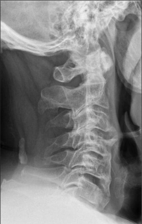
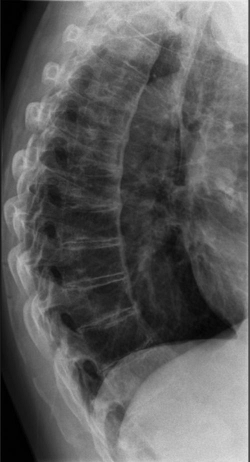
Our hospital performed X-rays of the cervical, dorsal, lumbar and pelvic spine which showed a calcification of the anterior common vertebral ligament with preservation of the disc spaces (Figs. 1 and 2).

Fig. 1.

Cervical spine X-ray: the height of the vertebral bodies are preserved. Calcification projected onto the soft tissues in the dorsal section in the occipito-vertebral ligament.

(0.04MB).
Fig. 2.

Dorsal spine X-ray: calcification of the common anterior vertebral ligament.

(0.04MB).

In view of these findings the patient was referred to the nuclear medical unit for a bone scan to rule out several causes of osteoarthritis, Forestier's disease among them.

Forestier's disease is related to abnormal bone growth.1–3 The main disease symptoms is calcification of the longitudinal ligaments of the spine, involving at least 4 contiguous vertebrae. Other criteria are the preservation of intervertebral disc spaces and the absence of inflammatory changes in intrapophysary or sacroiliac joints.4–6

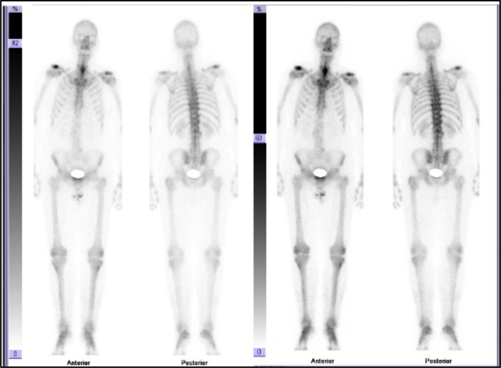
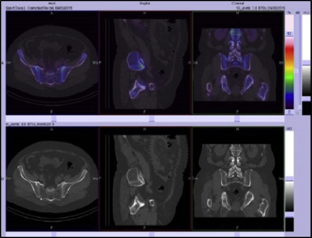
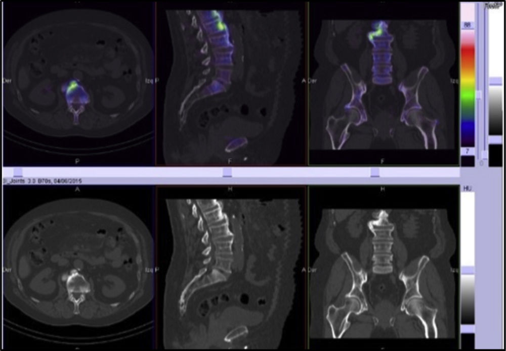
In the bone scan images there was no sign of articular pathological uptake (Fig. 3). Multimodality SPECT/CT tomographic images were then acquired (Figs. 4 and 5), which were used both for qualitative visual analysis of the sacroiliac joint, and for quantitative analysis which permitted the obtainment of indicators of the sacroiliac-promontory. In visual analysis calcification of the anterior common vertebral ligament was detected, together with absence of sacroiliac uptake. With this information and inferior promontory-sacroiliac indicators below the established cut-off level for diagnosis of sacroiliitis, diagnosis of ankylosing spondylitis was excluded, with support for the diagnosis of Forestier's disease.

Fig. 3.

Bone scan with normal pattern of uptake on the cervico dorsal skeleton and discrete uptake on the right-hand side of the lumbar rachis.

(0.05MB).
Fig. 4.

SPECT/CT slice images in axial, sagittal and coronal planes with low uptake of the radiopharmaceutical in sacroiliac joints. Sacroiliac/promontory values below cut-off level established for diagnosis of sacroiliitis.7,8

(0.07MB).
Fig. 5.

SPECT/CT: calcification of common anterior vertebral ligament and preservation of intervertebral disc spaces.

(0.06MB).
Ethical ResponsibilitiesProtection of people and animals

The authors declare that this research has not been carried out experiments on humans or animals.

Data confidentiality

The authors declare that they have adhered to the protocols of their centre of work on the publication of patient data.

Right to privacy and informed consent

The authors declare that no patient data appear in this article.

Conflict of interests

The authors have no conflict of interests to declare.

References
[1]
D. Hannallah, A.P. White, G. Goldberg, T.J. Albert.
Diffuse idiopathic skeletal hyperostosis.
Oper Tech Orthop, 17 (2007), pp. 174-177
[2]
J. Jensen, H. ur Rehman.
Diffuse idiopathic skeletal hyperostosis.
J Gen Intern Med, 8 (2013), pp. 31-32
[3]
F. Nascimento, L. Maranha, R. Oliver, H. Mello, Z. Demartini, G. Koppe, et al.
Diffuse idiopathic skeletal hyperostosis: a review.
Surg Neurol Int, 5 (2014), pp. S122-S125
[4]
D. Resnick, G. Niwayama.
Radiographic and pathologic features of spinal involvement in diffuse idiopathic skeletal hyperostosis (DISH).
Radiology, 119 (1976), pp. 559-568
[5]
P.D. Utsinger.
Diffuse idiopathic skeletal hyperostosis.
Clin Rheum Dis, 11 (1985), pp. 325-351
[6]
J. Forestier, J. Rotes-Querol.
Senile ankylosing hyperostosis of the spine.
Ann Rheum Dis, 9 (1950), pp. 321-330
[7]
L.S. Rodriguez, E. Ornilla, E. Prieto, M. Ribelles, M. Garcia-Velloso, J. Richter.
Quantitative bone SPECT/CT versus MRI in the early diagnosis of axial spondyloarthritis.
JNM, 54 (2013), pp. 1974
[8]
L. Sancho, E. Ornilla, E. Prieto, M. Ribelles, M. Garcia-Velloso, C. Caicedo, et al.
Diagnostic value of quantitative SPECT/CT versus MRI for the assessment of sacroiliitis in early axial spondyloarthritis. Diagnostic value of quantitative SPECT/CT versus MRI for the assessment of sacroiliitis in early axial spondyloarthritis.
Eur J Nucl Med Mol Imag, 40 (2013), pp. S242-S243

Please cite this article as: Morales Lozano MI, Ornilla E, Sancho L, Guillén EF. Single-photon emission computed tomography en el diagnóstico de un caso de enfermedad de Forestier. Reumatol Clin. 2019;15:307–308.

Copyright © 2017. Elsevier España, S.L.U. and Sociedad Española de Reumatología y Colegio Mexicano de Reumatología
Idiomas
Reumatología Clínica (English Edition)
Article options
Tools
es en

¿Es usted profesional sanitario apto para prescribir o dispensar medicamentos?

Are you a health professional able to prescribe or dispense drugs?