To study differences in demographic, clinical and immunologic characteristics, activity and cumulative organ damage according to age of onset in systemic lupus erythematosus (SLE).
MethodsCross-sectional study was performed including 204 SLE patients. Characteristics were compared between juvenile and adult-onset SLE patients using parametric and nonparametric tests (SPSS 23.0).
ResultsJuvenile-SLE patients had malar rash more often (78.9% vs 53%; p=0.001), oral ulcers (45.5% vs 17.5%; p=0.001), neurological involvement (13.1% vs 3.6%; p=0.02) nephritis (50% vs 33.9%), p=0.04) and haematological manifestations such as hemolytic anaemia (23.6% vs 5.4%; p=0.002) and leukopenia (46.1% vs 4.2%; p<0.001). Arthritis was more prevalent in adult-onset patients (70.9% vs 90%; p<0.04). Overall, 20% of juvenile patients had chronic damage (Systemic Lupus International Collaborating Clinics/Damage Index [SLICC/DI]≥1), However, the percentage of patients with irreversible damage was higher in the adult SLE patient group (24%, p=0.04). No statistically significant differences were found in other characteristics studied.
ConclusionIn summary, our study confirms the existence of differences in clinical manifestations, according to age at diagnosis of SLE. Juvenile-SLE patients showed a more aggressive clinical presentation.
Estudiar las diferencias en las características demográficas, clínicas, inmunológicas, en la actividad y daño crónico de acuerdo con la edad de aparición del lupus eritematoso sistémico (LES).
MétodosSe realizó un estudio de corte transversal incluyendo 204 pacientes con LES. Las características se compararon entre los pacientes con lupus de comienzo juvenil y del adulto utilizando testes paramétricos y no paramétricos (SPSS 23.0).
ResultadosLos pacientes con LES juvenil tenian más frecuentemente rash malar (78,9 vs. 53%; p = 0,001), úlceras orales (45,5 vs. 17,5%; p = 0,001), afectación neurológica (13,1 vs. 3,6%; p = 0,02) nefritis (50 vs. 33,9%, p = 0,04) y las manifestaciones hematológicas como anemia hemolítica (23,6 vs. 5,4%; p = 0,002) y leucopenia (46,1 vs. 4,2%; p < 0,001). La artritis era más frecuente en los pacientes con lupus del adulto (70,9 vs. 90%; p 0,04). El 20% de los pacientes juveniles tenían daño crónico (Systemic Lupus International Collaborating Clinics/Damage Index [SLICC/DI]≥1). Sin embargo,el porcentaje de pacientes con daño irreversible fue superior en adultos (24%, p = 0,04). No se encontraron diferencias estadísticamente significativas en relación con las otras características estudiadas.
ConclusiónEn resumen, nuestro estudio confirma la existencia de diferencias en las manifestaciones clínicas según la edad al momento del diagnóstico del LES. Pacientes con LES de comienzo juvenil mostraron una presentación clínica más agresiva.
Systemic lupus erythematosus (SLE) is a severe, multi-systemic rheumatic disease. It is most prevalent among women of childbearing age, but can occur in all ages. In about 10–20% of cases it begins before 16 years.1,2
Several studies have reported that age at onset has a modifying effect on disease expression. Juvenile-onset SLE (jSLE) tends to have a more aggressive presentation and course, high rates of organ involvement and increased need for long-term immunosuppressive medications.3–5
Most studies have found a higher prevalence of lupus nephritis and haematological involvement in jSLE. Among adults, mild forms prevail, being the discoid lupus and arthritis more common in this group. However, in respect of many other clinical manifestations, including neuropsychiatric lupus (NPSLE), serositis, and autoantibody profiles, reports are conflicting, which is probably due to the small number of patients analysed in these studies.4–7
These observations are of high importance since a high prevalence of severe organ involvement, as well as an early and prolonged exposure to high dose of steroid therapy and immunosuppressive drugs, raises concerns regarding the long-term morbidity and early mortality.
Although patients with jSLE present more often with severe forms of disease, differences in cumulative long-term damage, according to age of onset, is not well known. Few studies have proved the existence of a greater cumulative damage juvenile SLE patients. In 2008, Tucker and colleagues found a higher renal damage in these patients. In the same year, Ramirez Gomez and colleagues and Brunner and colleagues, showed that juvenile SLE patients had higher rates of activity (assessed by SLEDAI) but also may a higher chronic damage (assessed by SLICC/ACR index).8–10
The objectives of the study were to compare patients with juvenile-onset SLE with those with adult-onset subset, regarding demographic, clinical and immunologic manifestations and to compare the cumulative organ damage between the groups.
Patients and methodsCross-sectional study was performed.
Patients204 consecutive patients with diagnosis of SLE (according to the American College of Rheumatology 1997 criteria11), followed in our Rheumatology department of a tertiary University Hospital from Portugal, were included. Two groups were compared according to age of onset: juvenile and adult SLE. Juvenile-onset SLE was considered in those patients who began their disease at age of 16 or before.
No additional samples or outpatient attendances were required from patients for study purposes, thus ethical approval and informed consent were not required.
Clinical dataDemographic, clinical and immunological data were retrospectively obtained by consulting the clinical records and the national database Reuma.pt. The following variables were collected: gender, age at diagnosis, ethnicity, duration of follow-up, clinical manifestations including malar rash, discoid rash, photosensitivity, oral ulcers, arthritis, serositis, nephritis, neuropsychiatric involvement, haemolytic anaemia, leukopenia, thrombocytopenia and autoantibodies (antinuclear antibodies (ANA), anti-double stranded DNA antibody (anti-DsDNA), anti-Smith antibody (anti-Sm), lupus anticoagulant and anti-cardiolipin antibodies). Autoantibodies were considered positive if the value was above the cut-offs for the laboratory at least in one determination during the follow-up period, except for anti-cardiolipin antibodies, which were considered present if there was two positive doseaments apart from twelve weeks.
Disease activity was calculated using the Systemic Lupus Erythematosus Disease Activity Index (SLEDAI) and assessment of chronic organ damage was performed with the Systemic Lupus International Collaborative Clinics/American College of Rheumatology (SLICC/ACR) Damage Index. Both indexes were calculated in the last visit.
Statistical analysisDemographic, clinical and immunological variables were compared between two groups according to age at diagnosis: jSLE (≤16 years) and adult-onset lupus (>16 years).
Continuous variables are expressed as mean and standard deviation, if they have a normal distribution, or median and range, if distribution is highly skewed. Kolmogorov–Smirnov test was used to verify the normal distribution of continuous variables. Categorical variables are presented as absolute values and percentages.
For continuous variables, values were compared using Student's t-test test or Mann–Whitney test depending on normality distribution of variables. Categorical variables were analysed Fisher exact test.
The level of significance was set at 0.05. All statistical analyses were performed using SPSS, version 23.0.
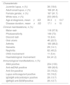
ResultsA total of 204 patients diagnosed with SLE were included, comprising 38 (18.6%) jSLE and 166 (81.4%) adult-onset SLE patients. 187 (91.7%) patients were female and had a mean age of 46.1±15.4 years and a mean of disease duration of 17.1±10 years (Table 1).
Demographic, clinical and immunological characteristics of the 204 SLE patients included.
| Characteristic | |
| Juvenile lupus, n (%) | 38 (18.6) |
| Adult-onset lupus, n (%) | 166 (81.4) |
| Female gender, n (%) | 187 (91.7) |
| White race, n (%) | 203 (99.5) |
| Age at diagnosis, mean±SD | 30.3±13.7 |
| Disease duration, mean±SD | 17.1±10 |
| Clinical manifestations, n (%) | |
| Malar rash | 118 (57.8) |
| Photosensitivity | 149 (73) |
| Discoid rash | 13 (6.4) |
| Oral ulcers | 46 (22.5) |
| Arthritis | 180 (88.2) |
| Serositis | 29 (14.1) |
| Nephritis | 82 (40.2) |
| CNS involvement | 11 (5.4) |
| Haematological involvement | 84 (41.2) |
| Immunological manifestations, n (%) | |
| ANA positive | 204 (100) |
| Anti-dsDNA positive | 189 (92.6) |
| Anti-Sm positive | 33 (16.2)) |
| Lupus anticoagulant positive | 33 (16.2) |
| IgG/IgM anticardiolipin positive | 28 (13.7) |
| IgM/IgG anti-B2GPpositive | 26 (12.7) |
SD, standard deviation; CNS, central nervous system; ANA, antinuclear antibodies; anti-DsDNA, anti-double stranded DNA antibodies, anti-Sm, anti-Smith antibody; anti-B2GP, anti-beta 2 glycoprotein.
The most prevalent clinical manifestations in this cohort of SLE patients were: arthritis (88.2%) and muco-cutaneous manifestations such as photosensitivity (73%) and malar rash (57.8%). The other systemic organ involvement was less common (Table 1).
Regarding immunologic profile, all patients had positive ANA and 92.6% had positive anti-dsDNA (Table 1).
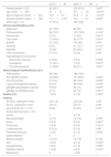
Comparison between juvenile and adult-onset SLE patients (Table 2) showed no statistically significant differences regarding demographic characteristic, except for age at diagnosis, as it was expected.
Comparison of demographic, clinical and immunological characteristics between 38 juvenile and 166 adult-onset Lupus patients.
| jSLE n=38 | aSLE n=166 | p | |
|---|---|---|---|
| Female gender, n (%) | 35 (92.1) | 152 (91.6) | 0.607a |
| Age (mean±SD) | 22±12 | 50.1±1.9 | <0.001b |
| Age at diagnosis (mean±SD) | 13.2±3.4 | 34.2±12 | <0.001b |
| Disease duration (mean±SD) | 17.1±11.56 | 15.1±9.7 | 0.962b |
| White race, n (%) | 37 (97.4) | 166 (100) | – |
| Clinical manifestations,n(%) | |||
| Malar rash | 30 (78.9) | 88 (53) | 0,002a |
| Photosensitivity | 29 (76.3) | 120 (72.8) | 0.418a |
| Discoid rash | 2 (5.2) | 11 (6.6) | 0.551a |
| Oral ulcers | 17 (45.5) | 29 (17.5) | 0.001a |
| Arthritis | 27 (70.9) | 153 (90) | 0.04a |
| Serositis | 8 (21) | 21 (12.7) | 0.141a |
| Nephritis | 19 (50) | 63 (33.9) | 0.04a |
| CNS involvement | 5 (13.1) | 6 (3.6) | 0,02a |
| Haematological involvement | |||
| Haemolytic anaemia | 9 (23.6) | 9 (5.4) | 0.002a |
| Leukopenia | 17 (46.1) | 7 (4.2) | 0.001a |
| Thrombocytopenia | 11 (28.9) | 45 (21) | 0.201a |
| Immunological manifestations,n(%) | |||
| ANA positive | 38 (100) | 166 (100) | – |
| Anti-dsDNA positive | 32 (84.8) | 157 (94.6) | 0.390a |
| Anti-Sm positive | 5 (13.2) | 28 (16.8) | 0.563a |
| Lupus anticoagulant positive | 5(13.2) | 28 (16.8) | 0.120a |
| IgG/IgM anticardiolipin positive | 5(13.2) | 23 (14) | 0.260a |
| IgM/IgG anti-B2GPpositive | 4 (10) | 22 (13) | 0.551a |
| Deaths,n(%) | 1 (2.6) | 2 (1.2) | 0.302a |
| Indexes | |||
| SLEDAI, median[min-max] | 2 [0–14] | 2 [0–23] | 0.39c |
| SLICC, median[min-max] | 0 [0–5] | 0 [0–5] | 0.09c |
| SLICC/ACR–DI ≥1, n (%) | 8 (20) | 40 (24) | 0.04a |
| SLICC/ACD domains, n (%) | |||
| Ocular | 0 | 2 (1.2) | – |
| Neuropsychiatric | 3 (7.9) | 12 (7.2) | 0.553a |
| Renal | 2 (5.3) | 9 (5.4) | 0.687a |
| Pulmonary | 1 (2.6) | 4 (2.4) | 0.201a |
| Cardiovascular | 4 (10.5) | 3 (1.8) | 0.08a |
| Peripheral vascular | 1 (2.6) | 3 (1.8) | 0.553a |
| Gastrointestinal | 0 | 0 | – |
| Musculoskeletal | 4 (10.5) | 13 (7.8) | 0.129a |
| Skin | 1 (2.6) | 2 (1.2) | 0.336a |
| Gonadal failure | 1 (2.6) | 3 (1.8) | 0.608a |
| Diabetes mellitus | 1 (2.6) | 4 (2.4) | 0.410a |
| Malignancy | 0 | 1 (0.6) | – |
Mann–Whitney test
Abbreviations: jSLE, juvenile-onset SLE; aSLE, adult-onset SLE; ANA, antinuclear antibodies; anti-DsDNA, anti-double stranded DNA antibody; anti-B2GP, anti beta2glyprotein; SLEDAI, Systemic Lupus Erythematosus Disease Activity Index S LICC/ACR-DI, Systemic Lupus International Collaborative Clinics/American College of Rheumatology Damage Index.
Juvenile SLE patients had more often muco-cutaneous manifestations, such as malar rash (78.9% vs 53%; p=0.001) and oral ulcers (45.5% vs 17.5%; p=0.001). Arthritis was more prevalent in adult-onset patients (70.9% vs 90%; p=0.04).
Neurological involvement was more commonly observed in jSLE group (13.1% vs 3.6%; p=0.02), as well as nephritis (50% vs 33.9%), p=0.04) and haematological manifestations such as haemolytic anaemia (23.6% vs 5.4%; p=0.002) and leukopenia (46.1% vs 4.2%; p<0.001).
Other manifestations, like photosensitivity, serositis and thrombocytopenia occurred in comparable percentages in both groups. Regarding immunologic profile, we did not find any statistically significant differences.
During the follow-up, the number of deaths was comparable in both groups. Were observed three deaths, two in the group of adults (one due to septic arthritis and septic shock and the other one in the context of pulmonary thromboembolism) and one in the group of juvenile-SLE (due to a severe pulmonary arterial hypertension). The mortality rate observed was 0.05 cases per 100 person-years in adult-onset SLE group and 0.15 in juvenile-SLE.
The disease activity score (SLEDAI) and the damage index SLICC/ACR were comparable in both groups. However, the percentage of the patients with irreversible damage (SLICC≥1) was superior in adult SLE patients group (24% vs 20%; p=0.04).
DiscussionThe study population consisted in patients from a public tertiary-level centre in Portugal. The clinical and demographic characteristic of the patients included in this study are similar to other Portuguese cohorts which were previously described.3,12
Consensus regarding the cut-off age for defining jSLE is still missing in literature. In our study, and also in most published studies, 16 years was considered the upper limit to define juvenile-Lupus. However, there are some studies that consider 18 years. Such variations may contribute to differences in results across studies.1,2
So far, few centres have deal with the comparison of paediatric and adult-onset lupus and only one meta-analysis has been published.4,7,13–15 In our study we found a higher prevalence of malar rash, oral ulcers, haemolytic anaemia, nephritis and CNS involvement in patients with juvenile onset, whereas arthritis was more prevalent in adult patients.
In fact, our data support the 2011 Livingstone meta-analysis results, where malar rash, nephritis, thrombocytopenia, haemolytic anaemia, seizure, fever, and lymphadenopathy were more common in jSLE whereas Raynaud's phenomenon and secondary Sjögren's syndrome were more prevalent in adult-onset group.13
Despite many controversies in the results of various studies, the most consistent among them is the higher prevalence of lupus nephritis. The most discordant results are with respect to CNS involvement probably because it is a rare manifestation and cohorts are too small, but we managed to prove in our cohort.13
Few studies have reported differences in chronic organ damage, which is one of the main concerns in the management of paediatric patients.8–10 In our study, SLICC/ACR damage index were comparable in both groups. However, the percentage of the patients with irreversible damage (SLICC≥1) was superior in adult SLE patients group. In fact, while some components of the SDI may be related to the use of corticoids (cataracts, avascular necrosis, osteoporosis, muscle atrophy/weakness) and cyclophosphamide (premature gonad failure, malignancy), some components may also be related to age (cataracts, diabetes mellitus, neoplasia, osteoporosis, stroke, and acute myocardial infarction) being expected a higher value in the adult group.
Yet it must be emphasised that 20% of paediatric patients had some chronic damage (SLICC/ACR DI≥1) which already shows a high percentage of morbidity in this group of patients.
Our study has a number of limitations. The use of a cross-sectional design, with retrospective analysis of medical records, may have underestimated the frequency of some clinical manifestation.
In summary, our study confirms the existence of differences in clinical phenotype, according to age at diagnosis of SLE. Although juvenile-SLE patients have high percentages of disease damage (20% of patients), we could not show differences comparing to adults groups, perhaps because many SLICC/ACR damage index items reflect age. It is clear that rheumatologists caring for children with SLE must be aware of the greater risk of major haematological, renal and CNS involvement as well as important long-term morbidity.
Ethical disclosuresProtection of human and animal subjectsThe authors declare that no experiments were performed on humans or animals for this study.
Confidentiality of dataThe authors declare that no patient data appear in this article.
Right to privacy and informed consentThe authors declare that no patient data appears in this article.
FundingThis research received no specific grant from any funding agency in the public, commercial, or not for-profit sectors.
Conflicts of interestThe authors declare that they have no conflict of interest.


