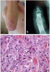
A 65-year-old Caucasian male consulted for the appearance of nodular skin lesions in form on firm, raised bumps with a red-brown and yellowish hue on the dorsal surface of the hands, forearms (Fig. 1a) and nasal region. He was followed in rheumatology outpatient clinic for 20 years with chronic erosive seronegative polyarthritis with erosions affecting mainly Distal Interphalangeal (DIP) joints (Fig. 1b). A combined therapy with methotrexate 20mg plus etanercept 50mg weekly was established with complete clinical remission at the onset of the lesions. A skin and metacarpophalangeal biopsy were performed. A proliferation of cells with a wide eosinophilic cytoplasm and the presence of multinucleated giant cells (Fig. 1c) was observed. The immunohistochemical analysis reveals that the cells express CD45, CD68, CD11b, and HAM-56, but not the S100 or CD1a protein, compatible with histiocytosis.
DiagnosisMulticentric reticulohistiocytosis (MRH) with joint and disseminated skin involvement was diagnosed.
EvolutionCutaneous lesions improved spontaneously in three weeks. Eighteen months later, only a few lesions were seen on the forearms and dorsum of the hands. Due to the spontaneous improvement and the absence of impact on the patient's life, no modification of therapy was considered, and the previous treatment was maintained. No another involvement was referred in the follow-up. A PET-scan was considered to rule out malignancy, but the patient died prior the scheduled date due a respiratory sepsis.
DiscussionMulticentric reticulohistiocytosis (MRH) is a rare idiopathic systemic inflammatory disease characterized by a tissue infiltrate of non-Langerhans histiocytic cells primarily in joints and skin but also affecting other organ systems.1 The disease often presents insidiously, beginning with articular symptoms and developing skin manifestations after a few months or even years. To our knowledge no report with such a long interval between articular and dermatological manifestations as in our patient (twenty years) has been described.2 This case, with an unusual lag between the presence of erosive joint involvement and the presence of skin lesions, highlights the importance of considering this entity in the differential diagnosis of patients with erosive joint lesions, predominantly in distal interphalangeal joints.
Authors’ contributionsAll authors contributed equally to the data collection, analysis, and writing of the manuscript.
Patient consentThe patient has provided informed consent for the publication of this case and the associated images.
Conflict of interestsThe authors declare that they have no competing interests.
We thank Dr. Carmen Fachal Bermudez from the Pathology Service of the Complexo Hospitalario Universitario de Vigo for her collaboration in the diagnosis of this case.


