


Systemic lupus erythematosus (SLE) is an autoimmune disease that causes multiple vascular complications, including endothelial cell damage. Nailfold capillaroscopy is the most effective non-invasive imaging technique for assessing the morphology of nailfold capillaries, and approximately half of the SLE patients have non-specific nailfold capillaroscopy abnormalities. Anti-uridin1-ribonucleoprotein antibodies are present in systemic lupus erythematosus patients with Raynaud's phenomenon, pulmonary artery hypertension, esophageal dysmotility, myopathy, and no severe renal involvement.
AimTo detect different patterns of nailfold capillaroscopic changes in SLE patients, their correlation with SLE disease activity, and anti-U1-RNP antibodies.
Patients and methodsA case–control study included eighty-six SLE patients, and disease activity was assessed using the SLEDAI-2K. All patients had a nailfold capillaroscopic examination. Anti-uridin1-ribonucleoprotein antibodies were measured in all patients.
ResultsAnti-uridin1-ribonucleoprotein antibodies had a significant inverse correlation with microhemorrhages and a significant direct relationship between anti-dsDNA antibody positivity and the presence of microhemorrhage. Additionally, a significant direct correlation was found between giant capillaries, venous plexus visibility, and higher ESR and CRP. Raynaud's phenomenon was significantly correlated with SLEDAI-2K, swollen joints, tender joints, and anti-dsDNA. Multiple linear regression analysis revealed that microhemorrhages and giant capillaries were the most significant predictors of lupus disease activity.
ConclusionOur findings highlight the prevalence of microvascular abnormalities in systemic lupus erythematosus, including tortuosity, crossing, elongation, microhemorrhages, and giant capillaries, emphasizing the importance of NFC in assessing microcirculation and disease activity. Also, it adds to the growing body of evidence supporting the prognostic value of capillary abnormalities, particularly microhemorrhages and giant capillaries, as predictors of disease activity in systemic lupus erythematosus patients. Nailfold capillaroscopic examination can assess lupus activity and potentially predict the risk of serious complications.
El lupus eritematoso sistémico (LES) es una enfermedad autoinmune que causa múltiples complicaciones vasculares, incluido el daño de las células endoteliales. La capilaroscopia del pliegue ungueal es la técnica de imagen no invasiva más eficaz para evaluar la morfología de los capilares del pliegue ungueal, y aproximadamente la mitad de los pacientes con LES tienen anomalías inespecíficas de la capilaroscopia del pliegue ungueal. Los anticuerpos anti-uridina1-ribonucleoproteína están presentes en los pacientes con lupus eritematoso sistémico con fenómeno de Raynaud, hipertensión de la arteria pulmonar, dismotilidad esofágica, miopatía y sin afectación renal grave.
ObjetivoDetectar diferentes patrones de cambios capilaroscópicos en el pliegue ungueal en los pacientes con LES, su correlación con la actividad de la enfermedad del LES y los anticuerpos anti-U1-RNP.
Pacientes y métodosUn estudio de casos y controles incluyó a 86 pacientes con LES y la actividad de la enfermedad se evaluó mediante el SLEDAI-2K. A todos los pacientes se les realizó un examen capilaroscópico del lecho ungueal. Se midieron anticuerpos anti-uridin1-ribonucleoproteína en todos los pacientes.
ResultadosLos anticuerpos anti-uridina1-ribonucleoproteína tuvieron una correlación inversa significativa con las microhemorragias y una relación directa significativa entre la positividad del anticuerpo anti-dsDNA y la presencia de microhemorragias. Además, se encontró una correlación directa significativa entre los capilares gigantes, la visibilidad del plexo venoso y una VSG y una PCR más altas. El fenómeno de Raynaud se correlacionó significativamente con SLEDAI-2K, articulaciones inflamadas, articulaciones sensibles y anti-ADNbc. El análisis de regresión lineal múltiple reveló que las microhemorragias y los capilares gigantes eran los predictores más importantes de la actividad de la enfermedad del lupus.
ConclusiónNuestros hallazgos resaltan la prevalencia de anomalías microvasculares en el LES, incluyendo tortuosidad, cruce, elongación, microhemorragias y capilares gigantes, enfatizando la importancia de NFC en la evaluación de la microcirculación y la actividad de la enfermedad. Además, se suma al creciente conjunto de evidencia que respalda el valor pronóstico de las anomalías capilares, en particular las microhemorragias y los capilares gigantes, como predictores de la actividad de la enfermedad en los pacientes con LES. El examen capilaroscópico del pliegue ungueal puede evaluar la actividad del lupus, y potencialmente predecir el riesgo de complicaciones graves.
Systemic lupus erythematosus (SLE) is an autoimmune disease that affects multiple systems and can lead to multiple vascular complications. SLE vascular disease causes endothelial cell damage and increases inflammatory mediators, leading to further dysfunction.1 Vascular complications associated with SLE include Raynaud's phenomenon (RP), microvascular and macrovascular thrombosis, pulmonary arterial hypertension (PAH), and inflammatory changes in blood vessels such as glomerulonephritis, cutaneous vasculitis, coronary vasculitis, cerebral vasculitis, alveolar capillaritis with pulmonary hemorrhage, and mesothelial vasculitis.2–4
Microcirculation is crucial for maintaining homeostasis by providing cells with oxygen, hormones, and nutrients and removing metabolic waste.5,6 Nailfold capillaroscopy (NFC) is the only most effective, non-invasive, easily repeatable imaging technique for analyzing the morphological assessment of nailfold nutrient capillaries. To date, there has not been a lot of consistent data on an “SLE-specific pattern.” This may be due to the complexity of SLE and its varying patterns of vascular involvement, which often manifest as capillary changes only during disease activity.7 Approximately 50% of SLE patients exhibit non-specific nailfold capillaroscopy abnormalities, including tortuous and meandering capillaries, bizarre loops, and prominence of the sub-capillary venous plexus.2,3 Gross abnormalities, such as giant capillaries or microhemorrhages, are less common.3
SLE patients with RP and pulmonary artery hypertension are more likely to have anti-uridin1-ribonucleoprotein (anti-U1-RNP) antibodies, which can also be associated with other symptoms of systemic sclerosis (SSc), including esophageal dysmotility, myopathy, and a reduced likelihood of severe renal involvement.2,8 Additionally, SLE patients exhibit higher levels of IgM anti-U1-RNP, while mixed connective tissue disease (MCTD) patients typically have positive IgG anti-U1-RNP but negative IgM anti-U1-RNP.9 These symptoms could indicate microcirculation problems, so evaluating microcirculatory abnormalities in SLE patients with anti-U1-RNP antibodies can aid in the early detection and treatment of internal organ involvement.
Based on this information, our study aimed to identify different patterns of nailfold capillaroscopic changes in SLE patients and examine their correlation with SLE disease activity and anti-U1-RNP antibodies.
Patients and methodsA case–control study was conducted on eighty-six SLE patients diagnosed according to the 2012 systemic lupus international collaborating clinics (SLICC criteria)10 after admission to our university hospital's rheumatology and rehabilitation department from December 2022 to December 2023.
Patients provided a complete history, including their age, gender, disease duration, present illness, drug use, age of disease onset, and family history.
The study excluded patients under eighteen years old and those with other autoimmune diseases such as mixed connective tissue disease, systemic sclerosis, dermatomyositis, or rheumatoid arthritis.
We excluded several factors that could affect NFC results. Patients with recent trauma or manual activities that might lead to microhemorrhages, or capillary damage were excluded. Additionally, those with poor nailfold hygiene, clubbing, congestive heart failure, smokers, caffeine users, and individuals unable to cooperate during the nailfold capillaroscopic examination were not included. We also excluded patients taking vasodilators, such as calcium channel blockers and phosphodiesterase inhibitors, which might decrease capillary visibility. Anticoagulants and antiplatelet medications, including aspirin, clopidogrel, and direct oral anticoagulants, were also reasons for exclusion due to their potential to increase microhemorrhages. Finally, patients using oral contraceptives or undergoing hormone replacement therapy were excluded because these could raise the risk of microthrombosis and vascular fragility.
Assessment of disease activityAll patients underwent clinical evaluation for SLE disease activity index 2000 (SLEDAI-2K) based on the presence of 24 descriptors in nine organ systems over the preceding 30 days.11 Lupus disease activity is classified into four groups: remission (SLEDAI-2K=0), mild (0<SLEDAI-2K≤6), moderate (6<SLEDAI-2K≤12), and severe (SLEDAI-2K>12).12
Nailfold video capillaroscopic examinationAll patients underwent NFC examination using the Dino-Lite Capillaroscopy Pro (MEDL4N Pro) at 200× magnification. Each patient's ring and little fingers on both hands were examined.5 Patients were asked to avoid caffeine and nicotine for 24h before the examination. NFC examinations were performed in a clinic room with a temperature controlled at 22–24°C, and vegetable oil was used to examine the digits.
Every nailfold image was divided into four consecutive 1mm fields and then evaluated for nailfold abnormalities such as13:
- (1)
The dominant capillary shape:
- (a)
Tortuous: Afferent and efferent limbs bend without crossing.
- (b)
Crossed: The afferent and efferent limbs cross each other.
- (c)
Elongated: Capillaries are elongated, with lengths exceeding 300μm.
- (2)
The capillary density: The average capillary number is 9–13 per 1mm.
- (3)
Microhemorrhages: Red or brown hemosiderin deposits next to the distal capillary row.
- (4)
Giant capillaries: Uniformly enlarged, with an apical diameter of ≥50μm.
- (5)
Neoangiogenic capillaries: Bushy, heterogeneous size and shape, and lack normal capillary loops.
- (6)
Avascular areas: A distance of 500μm or more between adjacent capillaries.
- (7)
Visible venous plexus: Vascular network at nailfold base.
Blood was drawn from the antecubital vein using 21 G needles and EDTA vacutainer tubes. The sample is divided as follows:
- (1)
Collect 1.8ml of blood in a tube with 0.2ml of trisodium citrate to detect ESR.
- (2)
Place 2ml of CBC in an EDTA-containing tube.
- (3)
3ml in a plain tube. Blood was left to clot in the incubator for 30min before being centrifuged for 15min. The expressed serum was used to measure serum creatinine, liver enzymes, serum albumin, CRP and ANA antibodies, anti-dsDNA antibodies, C3, C4, serum albumin, and anti-U1-RNP antibodies.
Data was collected and analyzed using SPSS Statistical Package, version 26 (SPSS Inc., Chicago, IL, USA, 1989–2019). Continuous data was expressed in mean±SD and range, while the nominal or categorical data were expressed in frequency (%). The relationships between nailfold capillaroscopic findings and clinical and laboratory characters in patients with systemic lupus erythematosus were studied and analyzed using Spearman correlation. The significance level considered was p≤0.05 (significant) or ≤0.01 (highly significant). One-way ANOVA was also applied for some continuous variables in the four categories of SLEDAI-2K. The capillaroscopic findings vs. SLEDAI-2K were treated in a multivariate sense by stepwise multiple regression, and the best equations with important characters for SLEDAI-2K prediction were estimated.
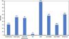
ResultsEighty-six SLE patients were included in the study. Their ages ranged from 18 to 58 years, with a mean age of (33.91±9.24). The mean disease duration among SLE patients was 6.52±5.15 years. Females comprised the vast majority of the study population (94.2%). Patients were divided into four groups based on SLEDAI-2K results: remission 16 (18.60%), mild 30 (34.88%), moderate 24 (27.91%), and severe 16 (18.60%).
One-third (31.39%) of SLE patients had swollen joints ranging from 1 to 16, with a mean of 3.96±3.91. The patients’ tender joints ranged from 1 to 10, with a mean of 4.24±2.4 and affecting 45 (52.33%) patients. Raynaud's phenomenon and ILD were observed in 43 (50%) and 3 (3.5%) of the patients, respectively, while ANA and anti-DsDNA antibodies were found in 84 (97.67%) and 49 (56.98%) of the patients. The patients’ 24-h protein levels in urine were divided into three categories: <150, <500, and ≥500, with corresponding percentages of 34 (39.53%), 30 (34.88%), and 22 (25.58%). Furthermore, approximately one-third of the SLE patients in the study (30.23%) tested positive for anti-U1-RNP antibodies, as shown in Fig. 1.

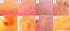
Each patient's nailfold capillaroscopic examination of the bilateral ring and little fingers yielded the following findings: 30 (34.9%) had tortuosity, 25 (29.4%) had crossing and elongation, 28 (32.6%) had microhemorrhages, 5 (5.8%) had giant capillaries, 6 (7%) had avascular areas, 7 (8.1%) had neoangiogenesis, and 29 (33.7%) had venus plexus visibility are shown in Fig. 2.

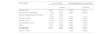
Table 1 enumerates the laboratory data of the SLE patients.
Baseline laboratory data of the studied SLE patients.
| Range | Mean±SD | |
|---|---|---|
| Hemoglobin (g/dl) | 7.6–14.0 | 11.267±1.494 |
| Platelets (103/μl) | 50.0–623.0 | 235.872±103.955 |
| Leukocytes (103/μl) | 2.2–11.5 | 5.302±2.079 |
| AST (U/l) | 8.0–261.0 | 27.252±28.579 |
| ALT (U/l) | 7.0–112.0 | 23.670±18.572 |
| Albumin (mg/dl) | 26.0–49.0 | 38.593±5.474 |
| Creatinine (μmol/l) | 22.0–143.0 | 61.206±20.192 |
| ESR (ml/dl) | 5.0–130 | 43.544±29.747 |
| CRP (mg/dl) | 0.1–36.0 | 7.657±9.117 |
| Complement 3 (mg/dl) | 0.09–1.8 | 1.011±0.445 |
| Complement 4 (mg/dl) | 0.01–0.6 | 0.223±0.122 |
CRP: c-reactive protein; ESR: erythrocyte sedimentation rate; AST: aspartate transaminase; ALT: alanine transaminase.
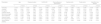
The study examined the correlation of capillaroscopic findings with age, disease duration, and clinical data in the SLE patients under study. The correlation of disease activity with neoangiogenesis, microhemorrhages, and avascular areas revealed a highly significant direct correlation (p=0.006, 0.026, and 0.005, respectively). The occurrence of RP showed a significant inverse correlation with capillary density (p=0.036), as well as a significant direct correlation with the incidence of microhemorrhages (p=0.033) and avascular areas (p=0.011). There was a significant direct correlation between the number of swollen joints and the visibility of the subpapillary venous plexus (p=0.025). Furthermore, there was a straightforward correlation between the number of tender joints and the extent of microhemorrhage (p=0.002; Table 2).
Correlation between the capillaroscopic findings and the age, disease duration, and clinical data in the studied SLE patients.
| Parameters | Age | Disease duration | SLEDAI-2K | Raynaud's phenomenon | Swollen joints | Tender joints | Interstitial lung disease | |||||||
|---|---|---|---|---|---|---|---|---|---|---|---|---|---|---|
| r | p-Value | r | p-Value | r | p-Value | r | p-Value | r | p-Value | r | p-Value | r | p-Value | |
| Tortuosity | −0.170 | 0.117 | 0.057 | 0.601 | 0.013 | 0.906 | −0.049 | 0.655 | −0.113 | 0.300 | 0.100 | 0.359 | −0.139 | 0.201 |
| Crossing | 0.207 | 0.057 | 0.014 | 0.901 | −0.062 | 0.570 | 0.033 | 0.761 | −0.050 | 0.651 | −0.072 | 0.514 | 0.016 | 0.881 |
| Elongation | 0.098 | 0.369 | 0.034 | 0.755 | 0.020 | 0.853 | −0.128 | 0.240 | −0.079 | 0.469 | −0.002 | 0.984 | 0.018 | 0.870 |
| Capillary density | −0.170 | 0.118 | −0.092 | 0.397 | −0.210 | 0.053 | −0.227* | 0.036 | 0.059 | 0.590 | −0.144 | 0.186 | −0.148 | 0.174 |
| Microhemorrhages | −0.069 | 0.526 | 0.004 | 0.970 | 0.241* | 0.026 | 0.230* | 0.033 | 0.125 | 0.253 | 0.326** | 0.002 | −0.029 | 0.789 |
| Giant Capillaries | −0.064 | 0.556 | −0.052 | 0.638 | 0.176 | 0.105 | 0.150 | 0.168 | 0.005 | 0.961 | 0.080 | 0.463 | −0.047 | 0.666 |
| Avascular areas | −0.140 | 0.198 | −0.120 | 0.272 | 0.303** | 0.005 | 0.274* | 0.011 | −0.020 | .854 | 0.147 | 0.175 | −0.052 | 0.634 |
| Neoangiogenesis | −0.070 | 0.521 | −0.139 | 0.203 | 0.293** | 0.006 | 0.211 | 0.051 | 0.022 | 0.839 | 0.113 | 0.299 | −0.057 | 0.605 |
| Venous plexus visibility | −0.012 | 0.914 | −0.007 | 0.946 | 0.153 | 0.159 | 0.203 | 0.060 | 0.241* | 0.025 | −0.009 | 0.938 | 0.109 | 0.316 |
Data are expressed as r (strength of correlation) and p (significance of correlation) where it is considered significant if ≤0.05 and highly significant if ≤0.01.
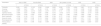
Table 3 examined the correlation between capillaroscopic findings and laboratory data from the SLE patients under study. The findings revealed that anti-U1-RNP antibodies had a significant inverse correlation with microhemorrhages (p=0.038) but no significant correlation with other capillaroscopic findings. In contrast, there is a significant direct relationship between anti-dsDNA antibody positivity and the presence of microhemorrhage (p=0.029). ANA antibodies and 24-h urine protein had no significant correlation with the capillaroscopic findings studied. Furthermore, a significant direct correlation was discovered between giant capillaries and venous plexus visibility and higher ESR (p=0.017 and 0.031, respectively) and CRP (p=0.005 and 0.003, respectively).
Correlation between the capillaroscopic findings and the laboratory data from the studied SLE patients.
| Parameters | Anti-U1-RNP | Anti-Ds-DNA | ANA | 24-h protein in urine | CRP | ESR | ||||||
|---|---|---|---|---|---|---|---|---|---|---|---|---|
| r | p-Value | r | p-Value | r | p-Value | r | p-Value | r | p-Value | r | p-Value | |
| Tortuosity | 0.049 | 0.651 | −0.103 | 0.345 | −0.049 | 0.655 | 0.038 | 0.730 | −0.054 | 0.621 | −0.112 | 0.304 |
| Crossing | −0.092 | 0.401 | 0.031 | 0.780 | 0.100 | 0.362 | −0.059 | 0.589 | −0.129 | 0.238 | −0.133 | 0.223 |
| Elongation | −0.087 | 0.426 | −0.013 | 0.908 | −0.071 | 0.515 | 0.141 | 0.196 | −0.129 | 0.238 | −0.065 | 0.552 |
| Capillary density | −0.066 | 0.547 | −0.096 | 0.381 | 0.098 | 0.370 | −0.012 | 0.915 | −0.184 | 0.089 | −0.131 | 0.229 |
| Microhemorrhages | −0.224* | 0.038 | 0.235* | 0.029 | 0.105 | 0.336 | 0.002 | 0.983 | −0.006 | 0.960 | 0.089 | 0.415 |
| Giant Capillaries | −0.164 | 0.133 | 0.117 | 0.285 | 0.038 | 0.726 | 0.177 | 0.103 | 0.303** | 0.005 | 0.258* | 0.017 |
| Avascular areas | −0.180 | 0.097 | 0.148 | 0.174 | 0.042 | 0.699 | 0.162 | 0.137 | 0.098 | 0.371 | 0.107 | 0.323 |
| Neoangiogenesis | −0.196 | 0.071 | 0.172 | 0.114 | 0.046 | 0.675 | 0.050 | 0.650 | 0.134 | 0.219 | 0.053 | 0.626 |
| Venous plexus visibility | −0.102 | 0.350 | 0.108 | 0.322 | −0.067 | 0.543 | 0.078 | 0.473 | 0.319** | 0.003 | 0.233* | 0.031 |
Data are expressed as r (strength of correlation) and p (significance of correlation) where it is considered significant if ≤0.05 and highly significant if ≤0.01.
CRP: c-reactive protein; ESR: erythrocyte sedimentation rate; ANA: antinuclear antibody; dsDNA: double strand deoxyribonucleic acid; U-1RNP: uridin1-ribonucleoprotein.
In Table 4, we investigated the relationship between anti-U1-RNP antibodies, Raynaud's phenomenon, and various clinical and laboratory parameters in SLE patients. Raynaud's phenomenon had a significant direct correlation with SLEDAI-2K, swollen joints, tender joints, and anti-dsDNA (p=0.001, 0.008, 0.033, and 0.001, respectively).
Correlation between the anti-U1-RNP antibodies, the incidence of Raynaud's phenomenon, and different clinical and laboratory parameters in studied SLE patients.
| Parameters | Anti-U1-RNP | Raynaud's phenomenon | ||
|---|---|---|---|---|
| r | p-Value | r | p-Value | |
| SLEDAI-2K | 0.068 | 0.535 | 0.338** | 0.001 |
| Interstitial lung disease | −0.125 | 0.251 | 0.063 | 0.562 |
| Raynaud's phenomenon | −0.051 | 0.643 | ||
| Swollen joints | −0.097 | 0.375 | 0.285** | 0.008 |
| Tender joints | −0.146 | 0.181 | 0.230* | 0.033 |
| 24-h protein in urine | 0.146 | 0.181 | 0.200 | 0.065 |
| ANA | 0.102 | 0.352 | 0.000 | 1.000 |
| Anti-dsDNA | 0.010 | 0.931 | 0.352** | 0.001 |
| Anti-U1-RNP | −0.051 | 0.643 | ||
Data are expressed as r (strength of correlation) and p (significance of correlation) where it is considered significant if ≤0.05 and highly significant if ≤0.01.
ANA: antinuclear antibody; dsDNA: double strand deoxyribonucleic acid; U1-RNP: uridin1riboneucleoprotein.
Multiple linear regression analysis was used to identify the most significant predictors of SLEDAI-2K in the nailfold capillaroscopic examinations of the SLE patients enrolled in this study. The results revealed that microhemorrhages (β=0.267, p=0.009) and giant capillaries (β=0.258, p=0.012) were the most significant predictors of lupus disease activity.
DiscussionSystemic lupus erythematosus (SLE) is an autoimmune disease frequently involving multiple vascular involvements. Impaired microcirculation and vascular hemodynamics could be early indicators of vascular disease.1 There has been an association between major capillary changes and the clinical course of SLE disease. Vascular pathologies in SLE, such as thrombotic events, microinfarcts, vasculitis, and perivascular inflammation, can cause various clinical symptoms. Patients with positive anti-U1-RNP antibodies who meet SLE criteria typically exhibit some clinical features commonly seen in MCTD patients, such as RP, lung damage, and changes in nailfold capillaroscopy.9 In the current study, we included 86 SLE patients. We conducted nailfold capillaroscopic examinations, which are considered the most reliable non-invasive method for analyzing microcirculation morphology in the nailfold area. These examinations focused on the bilateral ring and little fingers to identify various capillaroscopic patterns and their correlations with SLE disease activity and anti-U1-RNP antibodies.
Our findings revealed significant microvascular abnormalities in SLE patients; additionally, the scleroderma pattern in SLE patients can be detected without overlap syndrome with systemic sclerosis.14 The common morphological abnormalities that we identified in our study include tortuosity (34.9%), crossing (29.4%), and elongation (29.4%), which are consistent with the reported capillaroscopic abnormalities in SLE.15,16 Microhemorrhages were found in 32.6% of the studied patients, which is consistent with other studies showing microhemorrhages as a common capillaroscopic feature in SLE.7,15 Giant capillaries, which are uncommon (5.8%), were discovered in patients with severe disease activity (SLEDAI-2K), supporting previous research linking giant capillaries to scleroderma-like NFC patterns in severe cases.7,16
Our results also revealed no correlation between different capillaroscopic changes and the age and disease duration of the involved patients, which is consistent with various previous studies.17–19 Our study also discovered a direct weak correlation between disease activity and microhemorrhages, neoangiogenesis, and avascular areas, which is consistent with earlier research showing that such capillary changes are common in active SLE.3,18,20–22 These findings are most likely related to vascular destruction and neoangiogenesis, which are two major pathological processes in SLE. They occur due to vascular endothelial cell inflammation and endothelial cell damage caused by high levels of circulating autoantibodies during a disease flare. As a result, vascular endothelial growth factor (VEGF) and other mediators that promote vessel healing and neoangiogenesis are rapidly released. In SLE patients, circulating VEGF levels correlate with disease activity.21,22 The complex and multifactorial nature of autoimmune diseases could explain this weak correlation. Several factors can influence disease activity in autoimmune conditions, including immune system dysregulation, genetic predisposition, environmental triggers, and individual differences in disease presentation and progression. This complication may result in a lack of a direct and consistent link between disease activity and specific microvascular changes like microhemorrhages, neoangiogenesis, and avascular areas. Other confounding variables, such as treatment regimens, comorbidities, and patient-specific factors, may also influence the variability in the correlation between disease activity and microvascular changes. Further studies are needed to better understand the underlying mechanisms and factors driving this relationship.
Furthermore, a significant direct weak correlation was discovered between the number of tender joints and the presence of microhemorrhage, as well as a direct weak correlation between the number of swollen joints and the visibility of the subpapillary venous plexus, pointing out that swollen and tender joints can influence the visibility of the subpapillary venous plexus and microhemorrhage. Still, a weak correlation indicates that other variables may also affect this association.
The significant inverse weak relationship between the presence of RP and capillary density and a direct weak relationship with microhemorrhages and avascular areas is consistent with previous research demonstrating RP's impact on microvascular integrity in SLE patients.8,15,23 This correlation indicates that RP in SLE patients worsens microvascular damage, as evidenced by capillary loss and hemorrhages. This could be because RP is associated with non-specific NFC changes. The onset of RP and its impact on microvascular integrity may be caused by several factors, including endothelial dysfunction, immunological dysregulation, and inflammation. Furthermore, the observed weak associations between RP and these microvascular alterations may potentially be due to individual differences in illness severity and responsiveness to treatment. Another studies, however, found no significant differences between SLE patients with and without RP regarding avascular area and microhemorrhage.7,18
In terms of the correlation between the detected nailfold capillaroscopic changes and laboratory data in the studied SLE patients, our study discovered a significant inverse weak relationship between the presence of anti-U1-RNP antibodies and microhemorrhages, suggesting that anti-U1-RNP antibodies may protect against certain capillary changes, supporting this point, previous studies reported that patients with SLE pulmonary artery hypertension who had anti-U1-RNP antibodies had a higher 5-year survival rate and a better prognosis9,24; however, little is known about the effects of anti-U1-RNP antibodies on microvasculature. It is possible that the relationship between anti-U1-RNP antibodies and microhemorrhages is indirect, which explains the observed weak correlation. Still, other studies have found a link between anti-U1-RNP positivity and capillaroscopic abnormalities such as giant capillaries, avascular areas, and reduced capillary density.2 The difference in results could also be attributed to the wide range of anti-U1-RNP existence in SLE patients, which ranges from 3% to 69%.25 In contrast, there is a significant direct weak correlation between anti-dsDNA antibodies and microhemorrhages. The direct correlation between anti-dsDNA antibodies and microhemorrhages emphasizes the role of anti-dsDNA antibodies in causing vascular damage. These antibodies cause endothelial dysfunction and increased vascular permeability, resulting in capillary damage and microhemorrhages. Anti-dsDNA antibodies are frequently associated with more severe disease activity and complications, including lupus nephritis and cardiovascular risk. Microhemorrhages may be caused by complex, multifactorial mechanisms, such as complement activation, vascular inflammation, and secondary damage, rather than by anti-dsDNA antibodies alone, which could explain direct weak correlation between anti-dsDNA antibodies and microhemorrhages. Similar results between anti-dsDNA antibodies and hemorrhages were found in a previous study.19 Other studies found no significant differences in capillaroscopic findings between patients with positive and negative anti-dsDNA SLE.4,7 Our findings also revealed no significant correlation between ANA and capillaroscopic changes, which is consistent with previous research.18 This distinction highlights the varying effects of different autoantibodies on capillary changes in SLE.
Furthermore, we found a significant direct weak correlation, consistent with previous research,18,19 between the presence of giant capillaries, venous plexus visibility, and elevated ESR and CRP levels. Giant capillaries, venous plexus visibility, and increased ESR and CRP levels have a weak relationship that could be caused by a number of factors. Although CRP and ESR are systemic indicators of inflammation, they do not particularly represent microvascular anomalies that are localized. Giant capillaries or venous plexus visibility may indicate cumulative or chronic damage rather than acute inflammatory activity detected by ESR/CRP levels. Elevated ESR and CRP are markers of active inflammation, but they can also be influenced by factors unrelated to the microvascular changes under study, such as infection or comorbid inflammatory conditions. Our findings revealed no significant correlation between 24-h protein in urine and our examined capillaroscopic findings. These findings are consistent with those reported in previous studies.7,17 On the contrary, another recent study found a statistically significant correlation between capillaroscopic changes and 24-h protein levels in urine.19
Our results on the correlation between anti-U1-RNP and RP revealed no significant relationship; however, a previous study found a positive association between anti-U1-RNP and RP.26 Regarding the correlation between anti-U1-RNP and lupus disease activity, as measured by SELDAI-2K, our findings revealed no significant correlation, which is consistent with previous research. Similarly, there were no correlations between anti-U1-RNP and renal or joint involvement in SLE patients, which is consistent with previous research that found the prevalence of renal disorders and arthritis to be similar in both anti-U1-RNP positive and negative SLE patients.26,27 However, another study found an association between anti-U1-RNP and the occurrence of lupus nephritis; this could be attributed to the role of antigen–antibody interactions involving extractable nuclear antigens (ENAs), including RNP, in the pathogenesis of lupus nephritis28; however, a definitive relationship has not been established.
Our findings show a significant direct weak correlation between SLE patients with RP and positive anti-dsDNA antibodies, which is consistent with the previously reported findings.29 The fact that anti-dsDNA antibodies do not directly cause RP, but rather reflect systemic autoimmune activity, especially in SLE, may account for the weak correlation between SLE patients with RP and positive anti-dsDNA antibodies. Rather than being exclusively caused by these antibodies, RP can also be caused by endothelial dysfunction, localized microvascular damage, or vasospastic reactions. The findings of our study on renal involvement, as measured by 24-h protein in urine, revealed no significant correlation with the occurrence of RP in SLE patients, which is consistent with previous research.23 However, another study found that SLE patients with RP had glomerulonephritis.30 In terms of the observed significant weak correlation between RP and tender and swollen joints in SLE patients, previous literature reported the common occurrence of arthritis and/or arthralgia in patients with RP31; however, another study found that arthritis or arthralgia was equally present in SLE patients with and without RP.23 Since RP is primarily a vascular disorder caused by vasospasm and endothelial dysfunction, while tender and swollen joints are inflammatory joint manifestations linked to immune complex deposition, synovitis, and systemic inflammation, the weak but significant correlation between RP and these conditions in SLE patients can be explained by distinct pathophysiological mechanisms. It can restrict the correlation's strength.
Our study used multiple regression analysis to identify microhemorrhages and giant capillaries as significant predictors of disease activity in SLE patients. These findings are consistent with previous research demonstrating the predictive value of these capillaroscopic features in SLE, suggesting that nailfold capillaroscopy could be a useful prognostic indicator for lupus activity.19,20 For example, microhemorrhages were discovered to be a significant predictor of disease activity, indicating ongoing vascular damage and inflammation. Similarly, identifying giant capillaries as a predictor of disease activity lends credence to the idea that these capillary changes are indicators of severe microvascular involvement. Previous studies have shown that giant capillaries are associated with increased disease activity.20
Limitation and recommendationA large sample size is recommended for properly and accurately evaluating the various nailfold capillary changes in SLE patients. Future research should delve deeper into the distinct pathophysiological mechanisms underlying the various capillaroscopic abnormalities detected in SLE as influenced by different autoantibodies.
ConclusionOur study comprehensively evaluates NFC changes in SLE patients and their correlation with key clinical parameters such as anti-U1-RNP antibodies and disease activity. Our findings highlight the prevalence of microvascular abnormalities in SLE, including tortuosity, crossing, elongation, microhemorrhages, and giant capillaries, emphasizing the importance of NFC in assessing microcirculation and disease activity. Also, it adds to the growing body of evidence supporting the prognostic value of capillary abnormalities, particularly microhemorrhages and giant capillaries, as predictors of disease activity in SLE patients. NFC can assess lupus activity and potentially predict the risk of serious complications.
Ethical approvalThe study followed the Declaration of Helsinki's ethical principles after approval from our faculty's local research ethics committee (IRB No.: 17101701) and patients gave written informed consent to participate in this study. The study was registered in clinical trials “NCT05106998”.
FundingThe authors have no other relevant affiliations or financial involvement with any organization or entity with a financial interest in or financial conflict with the subject matter or materials discussed in the manuscript.
Conflict of interestsThe authors declare they have no conflict of interest.
We would like to express our deep gratitude to all patients who participated in the study.


