

We report the case of a 47-year-old patient who presented a bone neoplasia (T4N0 squamous cell carcinoma) located on the mandible, diagnosed in 2010 and initially treated with induction chemotherapy (cisplatin-5-fluorouracil in an Al-Sarraf protocol) and undergoing surgery, with bilateral neck dissection, hemimandibulectomy, left mandibular reconstruction with an fibula implant and skin grafting; subsequently he underwent treatment with 60 Gray of external radiotherapy and concomitant chemotherapy.
The patient continued treatment without noticeable adverse effects, while maintaining the correct function of the mouth and lower jaw.
A few months after the end of treatment he presented pain, swelling in the area of the jaw and progressive difficulty for flexion and extension of the neck and mouth opening.
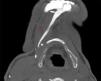
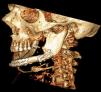
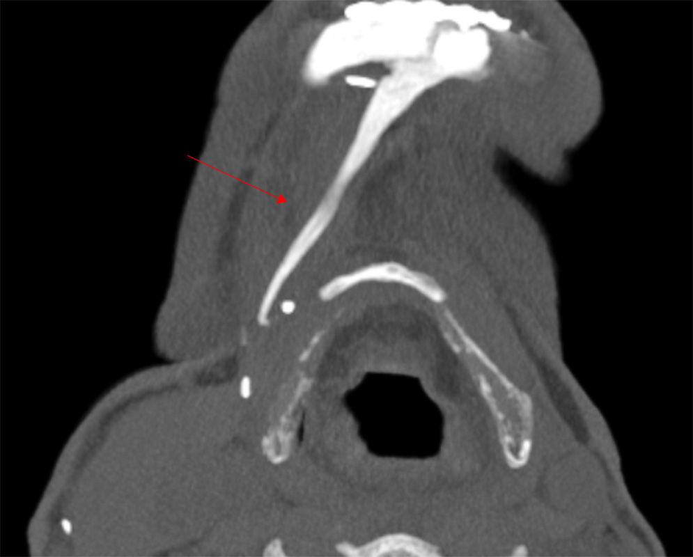
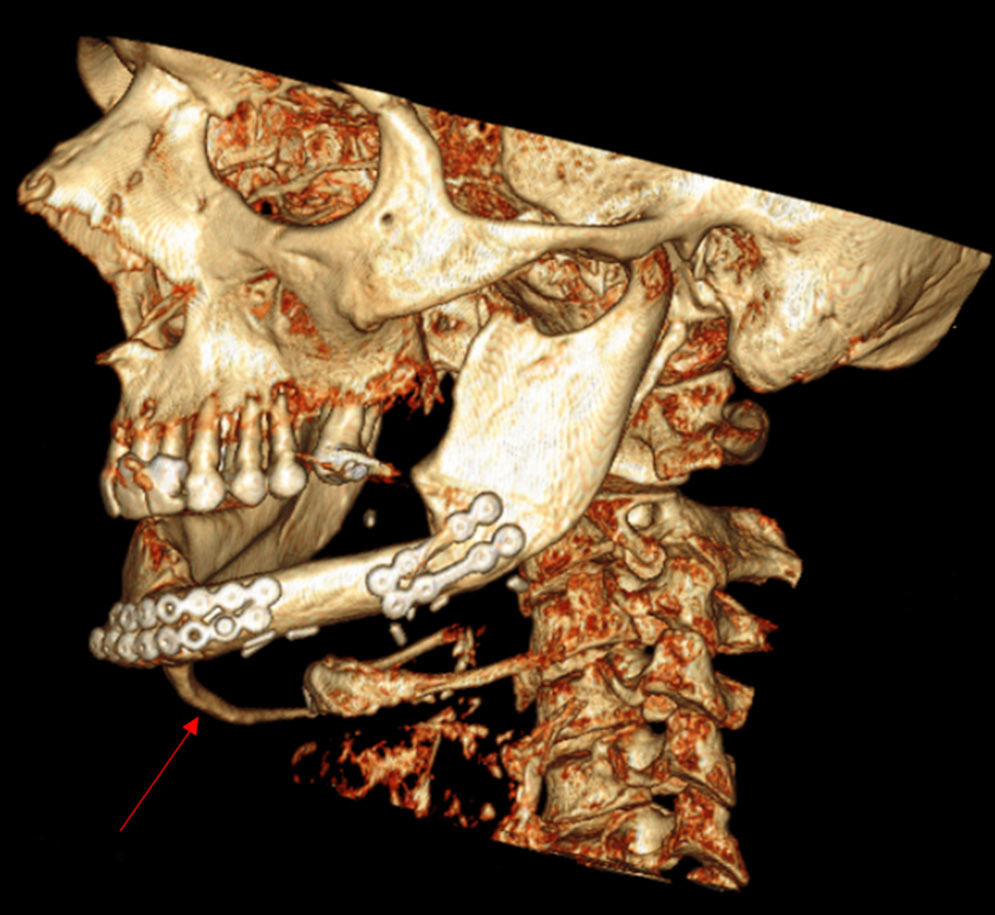
A bone scan was performed, showing localized technetium uptake in the mandible. An axial minimum intensity projection CT scan of the neck (MIP) (Fig. 1) and a 3D reconstruction (Fig. 2) were subsequently performed, showing an abnormal calcification linear stretching from the lower border of the mandible to the hyoid, following the anterior digastric muscle.
Circumscribed myositis ossificans appears during the second and third decades of life; the most common sites include the quadriceps muscle, leg, shoulder, arm and hand. Half of the patients have a history of multiple injuries or trauma in the affected area.1,2 A similar case of myositis ossificans in the facial muscles is described in the literature.3 Radiotherapy, surgery and prolonged immobilization of the mandible are factors that may play a role in the development of myositis ossificans in these cases.
Treatment is usually conservative; surgery is not recommended, especially in the early stages and in our patient was strongly discouraged due to recent radiotherapy. Treatment initially includes NSAIDs and rest followed by rehabilitation and physical therapy in order to restore function.4
Ethical ResponsibilitiesProtection of people and animalsThe authors declare this research did not perform experiments on humans or animals.
Data confidentialityThe authors declare that they have followed the protocols of their workplace regarding the publication of data from patients, and all patients included in the study have received sufficient information and gave written informed consent to participate in the study.
Right to privacy and informed consentThe authors have obtained the informed consent of patients and/or subjects referred to in the article. This document is in the possession of the corresponding author.
Conflict of InterestThe authors have no conflict of interest to state.
Please cite this article as: Pàmies A, Samitier A, Rodríguez-Fernández J, Fontova R. Miositis osificante en los músculos del cuello. Reumatol Clin. 2015;11:182–183.


