Journal Information
Vol. 8. Issue 4.
Pages 225-226 (July - August 2012)
Visits
43980
Vol. 8. Issue 4.
Pages 225-226 (July - August 2012)
Images in Clinical Rheumatology
Full text access
43-Year-Old-Male With a Right Pelvic Mass
Varón de 43 años con masa pélvica derecha
Visits
43980
Carmen Carrasco Cuberoa,
Corresponding author
maricarmen.carrasco@yahoo.es

Corresponding author.
, Josefa Jiménez Arjonab, Alfredo Michán Doñab
a Sección de Reumatología, Hospital Jerez de la Frontera, Cádiz, Spain
b Servicio de Medicina Interna, Hospital Jerez de la Frontera, Cádiz, Spain
This item has received
Article information
Full Text
Bibliography
Download PDF
Statistics
Figures (3)
Show moreShow less
Full Text
Clinical Case

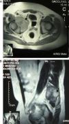
A 43-year-old male came to the clinic due to mechanical lower back pain, which had lasted for 2years. Physical examination showed painful limitation for right leg flexion and extension, with no neurological deficit. Laboratory analysis highlighted hemoglobin 10.6g/dl, normal ESR and GGT 111U/L (0–40). Simple X-ray (Fig. 1), CT (Fig. 2) and MR (Fig. 3) of the hips evidenced a 90mm×80mm×80mm mass in the right hemipelvis with an osteolytic component which affected half of the cotillus, iliac and pubic ramus, displacing the bladder and subjacent structures leftward.

Fig. 1.

Pelvic X-ray showing osteolysis (arrow) of the superior pubic ramus, with irregular bony margins and loss of cortical bone with no periosteal reaction.

(0.1MB).
Fig. 2.

Pelvis CT: lysis of the medial or superior portion of the acetabulum extended to the iliac and superior pubic ramus, associated with a large soft tissue mass measuring 12mm×74mm×73mm (arrow), infiltrating muscle.

(0.14MB).
Fig. 3.

(A and B) Axial and coronal MR of the hips RM in T2 sequence with contrast. A voluminous mass in the right hemipelvis is shown, affecting the anterior half of the cotillum, the iliac and pubic ramus, displacing the urinary bladder and adjacent structures leftward, with an isointense signal with bone and increased intensity with contrast, except in areas of intense necrosis.

(0.47MB).
Diagnosis and Progression

The histological study of an ultrasound guided biopsy concluded that the patient presented a peripheral primitive neuroectodermal tumor (PNET)/Ewing's sarcoma (ES). Chemotherapy was started with alternating cycles of vincristin/cyclophoshamide/adriamicine-iphosphamide/etoposid.

Discussion

Primitive neuroectodermic tumors are neoplasias with a differentiation to neuronal tissue which, if involving peripheral nerves, is called peripheral primitive neuroectodermic tumor.1 Because it shares the t(11;22) translocation with ES, it is considered the same disease. This tumor represents 3%–6% of solid tumors and 1.4%–1.8% of malignant processes, with an incidence of 3cases/million/inhab./year.2 90% of the cases appear between 5 and 30years and is more common in men. It is manifested by pain (in the pelvis, femur or humerus),3,4 swelling and, sometimes, fever, weight loss, anemia and leukocytosis.

X-ray findings reflect osteolysis, erosions, periostitis and soft tissue masses, as in our case. Immunohistochemistry is important to establish the differential diagnosis.5

Important findings in our patient were: age at presentation was uncommon as was the insidious nature of its progression, with no constitutional symptoms, fever or laboratory abnormalities. The natural history of PNET/ES is unknown although it tends to be aggressive in tumors developed in deep areas, when size is large or those presenting translocations.6 A combination of surgery, radiotherapy and chemotherapy attain an increase in survival and disease-free survival.7,8

References
[1]
O.P. Sangueza, P. Sangueza, L.R. Valda, C.K. Meshul, L. Requena.
Multiple primitive neuroectodermal tumors.
J Am Acad Dermatol, 31 (1994), pp. 356-361
[2]
M. Paulussen, B. Frohlich, H. Jurgens.
Ewing tumor: Incidence, prognosis and treatment options.
Paediatr Drugs, 3 (2001), pp. 899-913
[3]
J.P. Ginsberg, S.Y. Woo, M.J. Hicks, M.E. Horowitz.
Ewing's sarcoma family of tumors: Ewing's sarcoma of bone and soft tissue and the peripheral primitive neuroecctodermal tumors.
Principles and Practice of Pediatric Oncology, 4th ed.,
[4]
S.J. Cotterill, S. Ahrens, M. Paulussen, H.F. Jurgens, P.A. Voute, H. Gadner, et al.
Prognostic factors in Ewing's tumor of bone: Analysis of 975 patients from the European Intergroup Cooperative Ewing's Sarcoma Study Group.
J Clin Oncol, 18 (2000), pp. 3108-3114
[5]
C.S. Lee, M.C. Southey, H. Slater.
Primary cutaneous Ewing’ sarcoma/peripheral primitive neuroectodermal tumors in childhood. A molecular, cytogenetic and immunohistochemical study.
Diagn Mol Pathol, 4 (1995), pp. 174-181
[6]
M.L. Smith, H. Roberta.
Peripheral primitive neuroectodermal tumor presenting with diffuse cutaneous involvement and 7;22 translocation.
Med Pediatr Oncol, 30 (1998), pp. 357-363
[7]
S. Ferrari, E. Palmerini, M. Alberghini, E. Staals, M. Mercuri, E. Barbieri, et al.
Vincristine, doxorubicin, cyclophosfamide, actinomycin D, ifosfamide, and etoposide in adult and pediatric patients with nonmetastasic Ewing sarcoma. Final results of a monoinstitutional study.
Tumori, 96 (2010), pp. 213-218
[8]
J. Mora, C. De Torres, A. Parareda, F. Torner, P. Galván, E. Rodriguez, et al.
Treatment of Ewing sarcoma family of tumors with a modified P6 protocol in children and adolescents.
Pediatr Blood Cancer, 57 (2011), pp. 69-75

Please cite this article as: Carrasco Cubero C, et al. Varón de 43 años con masa pélvica derecha. Reumatol Clin. 2012;8:225–6.

Copyright © 2011. Elsevier España, S.L.. All rights reserved
Download PDF
Idiomas
Reumatología Clínica (English Edition)
Article options
Tools
es en

¿Es usted profesional sanitario apto para prescribir o dispensar medicamentos?

Are you a health professional able to prescribe or dispense drugs?