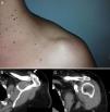
The patient was a 40-year-old woman. She came to the clinic due to the appearance of a tumor in the acromioclavicular joint (ACJ) after a vertical traction effort with the left arm (lifted, from the ground, a load of 40kg) 2 days prior. The lesion was painless, rounded and slightly compressible. It showed no inflammatory signs. There was no functional limitation of the shoulder but noticeable discomfort upon compressing the lesion (Fig. 1A).
Scanning with simple X-rays identified cortical irregularities on both sides of the ACJ. An ultrasound showed integrity of the rotator cuff and subacromial structures, with no alterations of the subacromial or subdeltoid bursae. There was acromioclavicular capsule distension and cortical marginal proliferation. The tumor was seen as a structure of defined edges, measuring 22mm×13mm and homogeneous anechoic content, compressible upon pressure. No continuity between the tumor and the joint capsule was found. A computed tomography (CT) described the injury as a rounded cystic structure dependent on the dorsal joint capsule which protruded cephalad (Fig. 1B and C).
Synovial cysts are evaginations synovium that occur as a result of an increase of synovial fluid pressure in the interior of a joint and persist in time; while the pressure difference between the joint capsule and the cyst is maintained or the communication channel between the two cavities is obliterated.1–3 Acromioclavicular cysts are rare and their presentation in the absence of rotator cuff disease is extremely rare.1,2 The only reported case-series of ACJ cysts dates from 2010 and described 5 cases in the presence of an intact rotator cuff.1 There are no reported cases since then. The rotator cuff tear leads to a homogenization of pressure in the subdeltoid and subacromial bursa. The latter transmits the increase in pressure on the acromioclavicular capsule, and should it be structurally weakened or anatomically conditioned, it will end up distended at the expense of the weaker wall usually in a dorsal or1,4–6 cephalad direction. From the radiological point of view, this flow of synovial fluid from the glenohumeral joint, to the subacromial bursa through the rotator cuff defect and reaching the ACJ, is known as the geyser sign and is demonstrable by arthrography or contrast MRI.6,7 Absent rotator cuff injury, the increase of pressure is usually secondary to extrinsic compression, for example, secondary to the vertical pull of the shoulder.
In the scant case reports, fistulization and acromioclavicular cyst recurrence are the main complications after needle aspiration,4,5,8,9 so this practice is not recommended unless there is clinical suspicion of an infectious process.10 The recommended treatment of these cysts is surgical removal of the tumor; the physician should be aware that, if case of a rotator cuff tear, repairs must be made before or during surgery.2,3,5,8,9 Sometimes, faced with a relapse, intervention should include removal of the distal clavicle.2,5,6
Ethical ResponsibilitiesProtection of people and animalsThe authors declare this research did not perform experiments on humans or animals.
Data confidentialityThe authors declare that they have followed the protocols of their workplace regarding the publication of data from patients, and all patients included in the study have received sufficient information and gave written informed consent to participate in the study.
Right to privacy and informed consentThe authors have obtained informed consent from patients and/or subjects referred to in the article. This document is in the possession of the corresponding author.
Conflict of InterestThe authors have no disclosures to make.
Please cite this article as: Guillén Astete CA, de la Casa Resino C. Quiste sinovial acromioclavicular con integridad del manguito rotador. Reumatol Clin. 2015;11:121–122.


