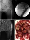
A 72 year-old woman with sequelae of multiple traumatisms in the left lower limb, in the form of loss of strength in all of the muscle groups. She visited the Emergency Department after low energy trauma, with the diagnosis of intertrochanteric femur fracture (Fig. 1A) Fig. 1, and she was received surgery with the emplacement of a short intermedullary nail (Fig. 1B). She was discharged from hospital after 7 days with no complications.
A. Simple X-ray of the left hip, anteroposterior projection, showing perthrocantheric fracture. B. Intraoperative fluoroscopy, anteroposterior projection, showing intramedullary nailing of the proximal femur. C. Simple X-ray of the left hip, anteroposterior projection, showing failure of the intramedullary nailing with luxation of the femoral head. D. Macroscopic appearance of the osteochondromatosis nodules sent to pathological anatomy.
6 weeks after surgery she visited the outpatient department with dysmetria of the limbs and a gradual increase in pain over recent days, without fever. The wound showed slight erythema with a slight increase in the local temperature. X-ray imaging showed failure of the nailing, with luxation of the femoral head (Fig. 1C), so that surgery was repeated.
During surgery capsular destruction was observed together with the presence of multiple tissue fragments with a hard elastic consistency and whitish-yellowish in colour (Fig. 1D). Suspecting infection, samples were taken for microbiology and anatomical pathology, and resection arthroplasty (Girdlestone) was performed. The sample cultures were negative, and anatomopathological study identified the samples as synovial osteochondromatosis.
Six months after the second operation the patient walked with great difficulty, using a raised insole to partially correct the dysmetria.
Synovial osteochondromatosis is a multifocal chondral metaplasia of the synovial tissue.1 The cartilaginous nodules which characterise it may detach from the synovial tissue, calcify and appear as free radio-opaque bodies in a simple X-ray.2 It may occur in any location, although it is more common in the large joints, and it rarely affects the hip.2 Dissemination outside the joint from a primary focus is exceptional,3 and it has been described as a possible cause of pathological fracture of the proximal femur.4
In patients diagnosed with the primary form and painful symptoms, the treatment is the surgical resection of the synovial tissue of the joint in question, using open or arthroscopic surgery.5
Conflict of interestsThe authors have no conflict of interests to declare.
Please cite this article as: García-Jiménez A, Álvarez A. Diagnóstico de osteocondromatosis como hallazgo casual en cirugía de fallo de enclavado entromedular de cadera. Reumatol Clin. 2020;16:125–126.


