We report the case of an 83-year-old man on rivaroxaban treatment, with pain in right shoulder, back of the upper arm, forearm and wrist, and inability to extend the wrist and fingers, as the result of an injury. The radiographs were normal, but thoracic CT showed an axillary artery pseudoaneurysm and a secondary haematoma that compressed the brachial plexus. This should be borne in mind in patients with painful shoulder, treated with anticoagulant therapy and without associated radiographic changes.
Se presenta el caso de un varón de 83 años en tratamiento con rivaroxabán, con dolor en hombro, cara posterior de brazo, antebrazo y muñeca derechos e incapacidad para extender muñeca y dedos, tras un traumatismo. Las radiografías son normales, pero en la TC torácica se objetiva seudoaneurisma de arteria axilar y un hematoma secundario que comprime el plexo braquial. Esta entidad ha de tenerse en cuenta en pacientes con hombro doloroso, anticoagulados y sin alteraciones radiológicas asociadas.
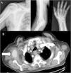
A male aged 83, receiving atrial fibrillation anticoagulation therapy with rivaxoaban, presented at the emergency department due to persistent pain his right shoulder which had spread to the back of his upper arm, forearm and wrist, of 10-day onset after an injury to the upper right limb in forced abduction. On examination a small throbbing axillary haematoma was observed, a strength of 1/5 on extending the fingers and of 3/5 on extending the wrist, hypesthesia in anatomical snuff box and first finger. Radial pulses were present and symmetrical. Requested X-rays showed no fractures or dislocations (Fig. 1a).
On suspicion of brachial plexus compromise cervical magnetic resonance imaging (MR) was requested, which resulted normal, together with electromyography which showed partial axonotmesis of the radial nerve. CT of the chest was also performed which showed an oval mass in the right armpit with irregular edges and a density similar to the axillary artery. As a result an angiography CTG scan was performed (Fig. 1b) which confirmed the said image was a continuity and pseudoaneurysm of the right axillary artery, axillary haematoma and axonotmesis of the ipsilateral radial nerve secondary to blunt trauma was diagnosed. The patient was referred for vascular surgery where a Doppler ultrasound scan was performed, draining of the haematoma and exclusion of the pseudoaneurysm through a stent. The patient is currently in physiotherapy treatment to recover strength lost from the injury and periodically goes to the outpatient rheumatology and vascular surgery department for check-ups.
DiscussionPost-traumatic pseudoaneurysm of the axillary artery is extremely rare, usually occurs in anterior dislocations of the shoulder and proximal fractures of the humerus. However, when it does occur, the brachial plexus is frequently damaged because it is in the same fascial compartment.1 On physical examination, the signs which should alert to a probable axillary pseudoaneurysm are: haematoma, throbbing mass, reduced or absent radial pulse, oedema of the affected limb, omalgia, brachial plexus nerve compromise and progressive dropping of haemoglobulin levels.2,3 In the case described the patient presented with most of these.
On occasion diagnosis of axillary artery pseudoaneurysm is complex due to latent neurological clinical features, depending on the compression of the perilesional haematoma and due to the fact that pulses may be retained, as was this case, from major collateral circulation of the upper limb.3 On suspicion of this we should request imaging tests such as the Doppler ultrasound scan, angiography CT or angiography MR. A further arteriography should be performed when the non-invasive tests are unable to lead to a diagnosis.4,5 In our case CT of the chest showed up the pseudoaneurysm.
Close follow-up of these patients should be made after intervention, to provide appropriate functional recovery of the brachial plexus, although complete recovery is not achieved in all cases.6 In this case the patient was reassessed one month after surgery, and presented with remission of pain, recovery of sensitivity but persistence of the inability to extend the fingers and wrist despite physiotherapy.
ConclusionPseudoaneurysm of the axillary artery is a possible cause of persistent shoulder pain where radiographs are normal and there is neurological compromise of the brachial plexus after an injury and this is more common in patients receiving anticoagulation treatment.
Conflict of interestsThe authors have no conflict of interests to declare.
Please cite this article as: Alcántara Fructuoso J, Bernal José L, Tornero Ramos C, Pretel Serrano L. Hombro doloroso en paciente anticoagulado con rivaroxabán. Reumatol Clín. 2020;16:120–121.


