

This study investigated the association between high-altitude residence (>2500m above sea level) and the presence of high probability of pulmonary hypertension (PH) in patients with systemic sclerosis (SSc).
MethodsA retrospective case–control study was conducted with 368 patients diagnosed with SSc at the rheumatology outpatient clinic of a university hospital in Bogotá, Colombia. Patients were divided into two groups based on the presence of high probability of PH. Clinical, demographic, and high-altitude residence data were collected and analyzed. A multiple logistic regression model was used to control confounding variables.
ResultsPatients residing at high altitudes had a significantly greater risk of presenting high probability of PH than those living at lower altitudes did (odds ratio: 2.0). Other significant factors included the diffuse cutaneous subtype of SSc and the presence of interstitial lung disease.
DiscussionHigh-altitude residence is a potential risk factor for presenting high probability of PH in SSc patients, warranting closer monitoring and tailored management in these populations. Further studies are warranted to confirm these findings.
Este estudio investigó la asociación entre la residencia en gran altitud (>2,500 metros sobre el nivel del mar) y la presencia de alta probabilidad de hipertensión pulmonar (HP) en pacientes con esclerosis sistémica (SSc).
MétodosSe realizó un estudio retrospectivo de casos y controles con 368 pacientes diagnosticados con ES en la consulta externa de reumatología de un hospital universitario en Bogotá, Colombia. Los pacientes se dividieron en dos grupos según la presencia de alta probabilidad de HP. Se recopilaron y analizaron datos clínicos, demográficos y de residencia a gran altitud. Para controlar las variables de confusión, se utilizó un modelo de regresión logística múltiple.
ResultadosLos pacientes que residían en gran altitud presentaron un riesgo significativamente mayor de presentar alta probabilidad de HP en comparación con aquellos que vivían a menores altitudes (Razón de Oportunidades: 2.0). Otros factores significativos incluyeron el subtipo cutáneo difuso de la ES y la presencia de enfermedad pulmonar intersticial.
DiscusiónLa residencia en gran altitud es un posible factor de riesgo para el desarrollo de alta probabilidad de HP en pacientes con SSc, lo que justifica una monitorización más estricta y un manejo personalizado en estas poblaciones. Se requieren más estudios para confirmar estos hallazgos.
Pulmonary hypertension (PH) occurs in 8–12% of patients with systemic sclerosis (SSc).1 PH accounts for approximately 30% of deaths related to SSc.2 PH in SSc can arise from several causes: pulmonary arterial hypertension or Group 1, due to arterial vasculopathy; PH due to left heart disease (Group 2); PH secondary to lung disease/hypoxia (Group 3); and PH secondary to veno-occlusive disease (Group 4).3 Group 5 PH corresponds to patients whose cause is unknown.
Several risk factors for developing PH in SSc patients have been identified, including lung fibrosis on computed tomography (CT), forced vital capacity (FVC)<80% of the predicted value, a history of Raynaud's phenomenon for at least three years before skin changes presented, limited cutaneous SSc (lcSSc), positive anti-centromere antibodies, and impaired carbon monoxide diffusion capacity (DLCO)<60%.4
High-altitude pulmonary hypertension (HAPH) arises from prolonged exposure to high altitudes and is recognized as a distinct category of PH according to the 2022 ESC and ERS classification.5 Populations residing at altitudes>2500 m above sea level (m.a.s.l., high altitude, HA) are at the highest risk for developing this condition.
The aim of this study was to determine whether HA residence was associated with the presence of high probability of PH in patients diagnosed with SSc from a rheumatology outpatient clinic in Bogotá, Colombia.
Materials and methodsStudy designThis is a retrospective, observational, analytical case–control study conducted with patients diagnosed with either established or early SSc who attended the outpatient rheumatology clinic at the Hospital Universitario Nacional (HUN), Bogotá, Colombia.
PatientsThis study was conducted in accordance with the ethical principles outlined in the Declaration of Helsinki and complied with all relevant national and international guidelines for research involving human participants. The research ensured that participants’ privacy and confidentiality were maintained throughout the study. Prior to the initiation of the study, ethical approval was obtained from the appropriate ethics committee. From January 2019 to December 2023, the medical records of patients attending the rheumatology clinic at HUN were reviewed for eligibility. Patients were eligible if they met the 2013 ACR/EULAR criteria for SSc or the 2011 VEDOSS criteria for early disease. Additionally, they must have had at least one echocardiogram report estimating the likelihood of PH and information regarding their residence during the preceding five years. Patients with polyautoimmunity (excluding Sjögren's syndrome), those with other non-SSc causes for PH, and those classified under PH Group 5 were excluded. Patients without a complete echocardiogram report or unclear residency data in the last five years were also excluded. A non-probabilistic convenience sampling method was used. A minimum sample size of 114 cases and 228 controls was estimated, considering an HA exposure probability of 0.6 and an estimated minimum risk with an odds ratio (OR) of 2.
Residence at high altitudePatients were considered to reside at HA if their places of residence in the last five years were all at an altitude>2500m.a.s.l. Those residing at altitudes<2500m were considered non-HA residents.
EchocardiographyThe included patients needed to have a complete echocardiogram report by a cardiology specialist. The echocardiograms must have been performed in geographic locations that matched the patient's altitude of residence.
High probability of pulmonary hypertensionPatients were considered to have a high probability of PH if the pulmonary artery systolic pressure (PASP) was>39mmHg, if the peak tricuspid regurgitation velocity (PTRV) was>3.4 m/s, or if the PASP was between 33–39mmHg with two additional echocardiographic signs of PH.3
Pulmonary hypertension groupsPatients were classified as having high probability of group 2 PH if, in addition to findings indicating a high probability of PH, clinical or echocardiographic findings of heart disease were present, such as systolic or diastolic heart failure, defined as a left ventricular ejection fraction (LVEF)<50%, segmental wall motion abnormalities, or type 2 or type 3 diastolic dysfunction. Additionally, they were considered to have a high probability of group 2 PH if they had a history of coronary artery disease or atrial fibrillation. Patients were classified as high probability of group 3 PH if, in addition to findings indicating a high probability of PH, there were signs of lung disease, such as interstitial lung disease (ILD) on chest CT, pulmonary function impairment with FVC<80% or DLCO<70%, chronic obstructive pulmonary disease (COPD) classified as moderate to severe, moderate to severe obstructive sleep apnea (OSA) or an indication for the use of positive pressure devices. As part of the analysis, ILD was considered severe if the extent was>20% or if the FVC was<70%. Finally, patients were classified as having high probability of group 4 PH if they had a history of chronic thromboembolic pulmonary disease or if compatible abnormalities were detected via ventilation/perfusion scintigraphy or angiography. Patients could be classified as both groups into high probability of PH groups 2 and 3. High probability of Group 1 PH was assigned only to patients with a high probability of PH who did not meet the criteria for any other group.
Case definitionPatients who met the criteria for a high probability of PH were classified as cases.
Control definitionPatients who did not meet the criteria for a high probability of PH were classified as controls.
Medical historyInformation was collected from the medical records registered in the reference hospital's system. Data on sociodemographic variables such as age and sex were extracted, along with clinical variables such as weight, height, past medical history, and prior pharmacological treatment, among others. Additionally, information on laboratory tests, diagnostic imaging, and pulmonary function tests were gathered.
Statistical analysisQuantitative variables are expressed as the means and standard deviations (SDs) or medians and interquartile ranges (IQRs). Categorical variables are expressed as frequencies and/or percentages. A bivariate analysis was conducted to assess differences between groups with and without high probability of PH, as well as between HA residents and non-HA residents. For categorical variables, Chi-square or Fisher's exact tests were used, as appropriate. For quantitative variables, the Mann–Whitney U test was performed.
A subgroup analysis was performed to determine the relationship between heart or lung disease and HA residence. Subsequently, HA residence and other risk factors were analyzed via a multivariate logistic regression model adjusted for both conventional risk factors for PH found in the literature and those identified in this study. ORs and 95% confidence intervals (95% CIs) were estimated. A p value of <0.05 was considered to indicate statistical significance. No data imputation was performed. Statistical analysis was performed with Stata 17® software.
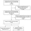
ResultsDuring the evaluation period, approximately 10,000 patients were seen in the rheumatology outpatient clinic at HUN. Among these patients, 410 had a diagnosis of SSc, but only 368 met the inclusion criteria. Ultimately, 114 cases and 254 controls were included in the analysis. A detailed description can be found in Fig. 1. According to the echocardiogram report, 114 patients (30.9%) were found to have a high probability of PH.
Sociodemographic characteristics
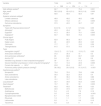
Table 1 presents the sociodemographic, clinical, and laboratory characteristics of the patients included in the analysis. Additionally, a comparison between the groups with and without high probability of PH is shown. Overall, 62% (n=231) of the patients were HA residents, with a higher prevalence among those with high probability of PH (75% vs 57%, p=0.001). Patients with high probability of PH were younger (56 vs 62 years, p=0.005). The sex distribution was similar in both groups, with >90% of the patients in each group being female. There were no statistically significant differences in sex distribution.
Description of patients based on the presence of high probability of pulmonary hypertension.
| Variable | Total | no PH | PH | p |
|---|---|---|---|---|
| n=368 | n=254 | n=114 | ||
| High altitude residenta | 62.7 | 57 | 75.4 | 0.001 |
| Age, yearsb | 58.2 (12.9) | 62.4 (13.1) | 56.3 (11.6) | 0.005 |
| Femalea | 92.6 | 92.5 | 92.3 | 0.87 |
| Systemic sclerosis subtypea | ||||
| Limited cutaneous | 48.9 | 49.2 | 48.2 | 0.86 |
| Diffuse cutaneous | 13.6 | 10.2 | 21 | 0.005 |
| Early/sine scleroderma | 37.5 | 40.5 | 30.7 | 0.07 |
| Symptoms | ||||
| Years with Raynaud phenomenonb | 8.6 (7.5) | 8.1 (7) | 9.7 (8.4) | 0.09 |
| Arthralgiaa | 45.5 | 43.4 | 50 | 0.24 |
| Dyspneaa | 41.3 | 28.3 | 70.1 | 0.005 |
| Dyspepsiaa | 63.7 | 58.9 | 74.3 | 0.005 |
| Clinical signsa | ||||
| Puffy fingers | 62.9 | 66.3 | 56.7 | 0.11 |
| Digital ulcers | 24 | 22.9 | 26.5 | 0.45 |
| Calcinosis | 17.7 | 17 | 19.3 | 0.59 |
| Telangiectasias | 61.5 | 57.5 | 70.5 | 0.19 |
| Tests | ||||
| Hemoglobin (mg/dl)b | 13.6 (1.7) | 13.7 (1.6) | 13.3 (1.7) | 0.03 |
| Antinuclear antibodiesa | 37.2 | 35.4 | 41 | 0.3 |
| Anti-centromere antibodiesa | 65 | 66.9 | 60.7 | 0.25 |
| Anti-Scl-70a | 9.6 | 9.5 | 9.8 | 0.9 |
| Interstitial lung disease in chest computed tomographya | 39 | 33.1 | 51.3 | 0.001 |
| Severe Interstitial lung disease in chest computed tomographya | 13.7 | 10.3 | 20.9 | 0.009 |
| Forced vital capacity<80%a | 23.8 | 20.4 | 29.7 | 0.11 |
| Pulmonary artery systolic pressure (mmHg)b | 36 (18.2) | 25.5 (10.5) | 51.3 (14.7) | <0.001 |
| Capillaroscopy patterna | ||||
| Nonspecific | 79.4 | 79.3 | 79.6 | 0.96 |
| Early scleroderma | 30.3 | 32.2 | 25.9 | 0.4 |
| Active scleroderma | 36 | 36.3 | 35.2 | 0.88 |
| Late scleroderma | 13.1 | 10.7 | 18.5 | 0.16 |
| Lung diseasea | 40.7 | 34.6 | 54.3 | 0.005 |
| Heart diseasea | 15.7 | 39 | 50 | 0.005 |
| Treatmenta | 62.2 | 60.2 | 66.6 | 0.23 |
| Methotrexate | 44.5 | 47.6 | 37.7 | 0.07 |
| Azathioprine | 5.4 | 4.3 | 7.8 | 0.16 |
| Cyclophosphamide | 3.5 | 0.8 | 9.6 | <0.001 |
| Mycophenolate mofetil | 6.7 | 5.9 | 8.7 | 0.31 |
| Rituximab | 1.3 | 1.1 | 1.7 | 0.66 |
| Leflunomide | 0.2 | 0 | 0.8 | 0.13 |
| Tacrolimus | 0.2 | 0.3 | 0 | 0.5 |
PH, high probability of pulmonary hypertension.
The most common form of SSc was lcSSc (48.9%), followed by the early/sine scleroderma subtype, with diffuse cutaneous SSc (dcSSc) being the least common subtype. There was a greater number of patients with dcSSc among those with high probability of PH (p=0.005).
Symptoms and signsNo differences were found in the number of years with Raynaud's phenomenon or joint involvement. In the group of patients with high probability of PH, dyspepsia and dyspnea were more common (74% vs 58% and 70%vs28%, respectively, p<0.05). No differences were found between the groups with and without high probability of PH in terms of the frequency of puffy fingers, digital ulcers, calcinosis, or telangiectasias. With respect to missing data, 25% of patients had no recorded information about the presentation of puffy fingers.
Paraclinical characteristicsThe hemoglobin concentration was greater in patients without high probability of PH (13.7 vs 13.3mg/dL, p=0.03). No differences were found between the groups regarding positivity for antinuclear antibodies (ANAs), the ANAs centromere pattern, or the presence of anti-Scl-70 antibodies. The presence of ILD and extensive ILD on chest CT was more common in patients with high probability of PH (51% vs 33%, p=0.001, and 20% vs 10%, p=0.009, respectively). Eighty percent of patients had no information regarding chest CT. There were no statistically significant differences in the frequency of patients with a FVC<80%. Thirty percent of patients had no information on spirometry. The PASP was greater in patients with high probability of PH (51 vs 25mmHg, p=0.001).
Nailfold capillaroscopyThe most common pattern on nailfold capillaroscopy (NFC) was the active scleroderma pattern (36%), followed by the early scleroderma pattern (30%), and finally the late scleroderma pattern (13%). No significant differences were found between the groups with and without high probability of PH. Fifty-three percent of patients had no information regarding NFC.
Pulmonary hypertension groupsAmong the patients with high probability of PH, 50% (n=57) were classified into high probability of group 2 PH, and 54% (n=62) were classified into high probability of group 3 PH. A total of 46% (n=52) were classified as high probability of group 1 PH. None of the patients had high probability of group 4 PH.
Cardiac and respiratory comorbiditiesPatients with high probability of PH had a higher frequency of pulmonary and cardiac comorbidities (54% vs 34% and 50% vs 39%, respectively).
Pharmacological treatmentOverall, 62% (n=229) of the patients were receiving some form of pharmacological treatment for SSc. There were no differences between the groups with and without high probability of PH. However, the use of cyclophosphamide was more common in the high probability of PH group (9.6% vs 8%, p=0.001).
When patients were compared based on whether they were HA residents or not (Supplementary Table 1), a greater percentage of patients with high probability of PH were found in the HA resident group (37% vs 20%, p=0.001). Additionally, HA residents had a greater percentage of dyspepsia (68% vs 55%, p=0.019), puffy fingers (68% vs 45%, p=0.001), telangiectasias (66% vs 52%, p=0.009), extensive ILD (16% vs 9%, p=0.001), and treatment with cyclophosphamide (5% vs 0%). They also had higher hemoglobin levels (13.9 vs 13.1mg/dL, p=0.001) and greater positivity for ANAs (42% vs 28%). The non-HA resident group had a greater percentage of patients with abnormal NFC patterns (89% vs 74%, p=0.01) and early scleroderma patterns (47% vs 21%), a greater percentage of patients receiving pharmacological treatment for SSc (72% vs 56%), and a greater frequency of methotrexate use (8% vs 5%). No differences were found in the frequency of cardiac or pulmonary diseases when these groups were compared.
Bivariate and multivariate analysisConsidering the differences between groups, a bivariate and multivariate analysis was performed to study the factors associated with high probability of PH, adjusted for variables identified in this study and previously recognized: sex, age, dcSSc, presence of anti-centromere antibodies, anti-Scl-70 antibodies, joint involvement, gastrointestinal symptoms, telangiectasias, puffy fingers, and cyclophosphamide use (Supplementary Table 2). The variables of anti-centromere antibodies and severe pulmonary disease were excluded from the multivariate logistic regression model because of the absence of significant differences in the bivariate analysis. We found that being an HA resident was associated with the presence of high probability of PH of any type, even after adjusting for other factors (OR 2.0, 95% CI: 1.16–3.5, p=0.012). Furthermore, it remained a risk factor for high probability of PH even after adjusting for pulmonary disease (OR: 2.4, 95% CI: 1.2–5.1, p=0.001), cardiac disease (OR: 2.6, 95% CI: 1.3–5.2, p=0.004), and even the combination of these two conditions (OR: 2.8, 95% CI: 1.4–5.4, p=0.003). The combination of being an HA resident and having pulmonary disease further increased the risk of high probability of PH (OR: 3.38).
DiscussionThis study revealed a significant association between residence at HA and high probability of PH, which persisted even after adjusting for confounding variables. Previous studies have reported a prevalence of PH between 5% and 23% in populations living above 3200m.a.s.l. in the Altiplano of South America.6–8
SSc was more frequently observed in women. A systematic review revealed that the pooled prevalence in women was 28/100,000, whereas it was 6/100,000 in men.9 Male sex has been described as a poor prognostic factor in the presentation of SSc. One study indicated that the five-year survival rate for men was 73%, whereas it was 45% for women at ten years.10 In this study, no relationship was found between sex and the presentation of high probability of PH, as in previous reports.11,12
Regarding the subtypes of SSc, a previous association between dcSSc and PH has been reported.2 Patients with dcSSc exhibit a more severe vasculopathic phenotype, which is likely related to a higher frequency of PH.13
A study in China by Huang et al.14 found that SSc patients with PH had more digital ulcers, gastroesophageal reflux, and telangiectasias than did those without PH. In our study, when multivariate analysis was performed, only the presence of telangiectasias and gastroesophageal reflux were associated with the presentation of SSc-related high probability of PH.14 In contrast to these findings, our study revealed that HA residents who presented high probability of PH had a greater frequency of gastrointestinal symptoms and presented more frequently with puffy fingers and telangiectasias. However, after multivariate analysis was performed, the associations between high probability of PH and these variables were not maintained.
Patients with SSc have a greater cardiovascular risk associated with heart failure and pulmonary congestion,15 which is consistent with what was identified in this study, where cardiac disease is associated with high probability of PH. A systematic literature review by Chen et al. revealed increased risks of stroke, cardiovascular disease, acute myocardial infarction, and venous thromboembolism.16 Our findings indicate that the impact of cardiac disease in HA residents might be greater than that in non-HA residents.
ILD is common in patients with SSc and is also a cause of PH. Xanthouli et al. conducted a study on SSc and PH, describing the presence of ILD in 26.2% of cases.17 Our study revealed that the proportion of HA residents with high probability of PH with ILD was 39.7%, which was not significantly different from that of non-HA residents.
Anemia is a well-known prognostic factor for patients with SSc. Xanthouli et al., in their cohort of SSc and PH patients, reported a hemoglobin level of 13.39±1.37g/dL. In this study, HA residents with a high probability of PH had an average hemoglobin level of 13.9g/dL, which was significantly different from that of non-HA residents.17
Positive ANAs in SSc patients are associated with greater organ involvement and vasculopathy, including PH.18 In this study, we found no difference in the frequency of ANA between groups.
Anti-centromere and anti-Scl-70 antibodies are associated with cardiopulmonary complications in this disease. Anti-centromere antibodies are linked to PH and are not related to ILD and are associated with an increase of 2.5mmHg per year.18 These antibodies were positive in 65.7% of HA residents. Similarly, the presence of anti-Scl-70 antibodies is associated with ILD and the development of PH. In the present study, this antibody was found in a low proportion.
Compared with the cohort of Xanthouli et al., who reported a PASP of 23.01±10.35mmHg in their cohort, the group of HA residents with SSc and high probability of PH had an average PASP of 36.5 (19.9)mmHg.17
Pulmonary function alterations suggestive of PH (FVC/DLCO ratio>1.8) were observed in 15 patients, of whom only 8 (53.3%) were in the high probability of PH group. According to Yuer Li et al., the FVC/DLCO ratio is a predictor of PH in patients with SSc, where an FVC/DLCO ratio>0.44 identifies patients with PH and is an independent predictor of 5-year all-cause mortality.19 The data presented on pulmonary function and tomography findings were included for descriptive purposes only, as the lack of data in a significant number of patients limits their interpretation. In a future study, a broader inclusion of these variables may be relevant.
An association between progressive capillary loss and progression to an active or late scleroderma pattern in NFC and the development of incident PH has been reported.20 In our study, an abnormal NFC pattern was observed in 74.1% of patients, with an early scleroderma pattern in 21.5%, an active pattern in 38.7%, and a late pattern in 13.7%.
The optimal medical treatment for HAPH involves a multimodal approach. As reported by Li et al.,21 it is recommended that patients experiencing pulmonary vasoconstriction due to low oxygen tension migrate to altitudes<2500 m.a.s.l.22 With respect to pharmacological management, data from randomized clinical trials are scarce; pulmonary vasodilators such as phosphodiesterase-5 inhibitors are recommended.21,23–25 Unfortunately, information about treatment for PH was not available in the records of our patients.
Limitations of this study include the inability to establish causality; however, our data suggests an association between residence at HA and PH. Another limitation is the inclusion of patients who did not undergo right heart catheterization (RHC) because of the nature of the study. Nonetheless, depending on the patient's clinical context, RHC may not be strictly necessary.26 This is particularly relevant in resource-limited settings such as ours, where logistical, technical, and economic constraints limit the use of some diagnostic tools considered the gold standard. Additionally, echocardiography has shown acceptable diagnostic performance for PH.27 For this reason, PH frequency estimates via echocardiography have been used in previous studies, in countries such as the United States,26 the Netherlands,28 Italy,29 and Australia.30 Nevertheless, studies confirming these findings in patients with results of RHC are warranted,
Finally, we acknowledge another limitation: the lack of data on smoking status, which has been identified as a factor associated with PH, particularly in relation to COPD development.
This study revealed that individuals meeting the criteria for SSc or those in the early stages of SSc who reside at HA have an association with the presence of high probability of PH. Given the results of this research, these individuals should undergo more stringent screening and potentially have shortened follow-up intervals.
Ethical considerationsThis study was approved by the local Ethics, Bioethics, and Research Committee.
CRediT author statementLuis Javier Cajas: Conceptualization, Methodology, Investigation, Data Curation, Formal analysis, Resources, Writing—original draft, Writing—review & editing, Supervision, Project administration. Julia Recalde Reyes: Conceptualization, Investigation, Writing—original draft. Javier Alejandro Correa: Conceptualization, Investigation, Writing—original draft. Wilder Carvajal: Conceptualization, Resources, Writing—original draft, Writing—review & editing. Carolina Torres: Conceptualization, Resources, Writing—original draft, Writing—review & editing. José S. Cortés: Conceptualization, Resources, Writing—original draft, Writing—review & editing.
FundingSelf-funded.
Conflict of interestNone to declare.
The followings are the supplementary data to this article:


