


A 65 year-old female with a history of sarcoidosis with pulmonary and joint involvement, who after 5 years of diagnosis begins with central nervous system involvement manifesting as diplopia. She presents normal analysis results. In imaging results, a mass is identified in the right intraconal space; it depends of right optic nerve, and shows multiple lymph node involvement. Biopsy was performed diagnosed with large B-cell lymphoma, an atypical form of tumor associated with sarcoidosis.
Mujer de 65 años de edad con antecedentes de sarcoidosis, con afectación pulmonar y articular, que tras 5 años del diagnóstico comienza con afectación del sistema nervioso central, manifestándose como diplopía. Presenta analíticas normales. En las pruebas de imagen se identifica masa intraconal derecha dependiente del nervio óptico derecho, así como múltiple afectación adenopática. Se realizó biopsia con diagnóstico de linfoma B de células grandes, forma atípica de tumor asociado a sarcoidosis.
Sarcoidosis is characterized by the accumulation of T cells and macrophages, which form noncaseating epithelioid granulomata. The involvement of lung parenchyma, joints and uveitis is also characteristic.1 The development of a lymphoma in the setting of sarcoidosis is exceptional and, thus, we consider it interesting to report a new case in which, it presented as an eye mass.
Clinical ObservationThe patient was a 65-year-old woman with no medical history of interest. She had been diagnosed 5 years earlier with sarcoidosis, showing hilar lymph node involvement, with granulomata in the transbronchial biopsy, and had received small doses of steroids. Four years later, she presented with pulmonary progression (chest radiography, high-resolution computed tomography), with normal respiratory function tests, and received no treatment. One year later, she developed right eye pain, intermittent diplopia and asthenia. The ocular fundus, lumbar puncture and general examination were normal, except for a small change in left supraclavicular lymph node. Magnetic resonance imaging revealed a lesion measuring 11mm×17mm×16mm, with well-defined borders, dependent of the optic nerve, did not infiltrate muscle tissue and attracted gadolinium. There was another lesion of less than 1cm in left masticatory space with the same characteristics (Fig. 1). Thoracoabdominal pelvic computed tomography discovered enlarged multiple mediastinal lymph nodes and iliac chains, and 18F-fluorodeoxyglucose-positron emission tomography/computed tomography (PET/CT) showed hypermetabolic lesions at the eye level, the left masticator space, and sacral and thoracic vertebral lymph node involvement. A biopsy of the supraclavicular lymph node demonstrated a diffuse large B cell lymphoma with a post-germinal center immunophenotype. Cytometry recognized an associated T lymphoid population that expressed CD3+, CD5+, CD8+, CD4− and a high index of proliferation (Ki67). Using B- and T-cell rearrangement studies, we confirmed a monoclonal B population. Although a biopsy of the iliac crest gave no sign of lymphoma, we did find evidence with granulomatous infiltration with multinucleated giant cells.
Discussion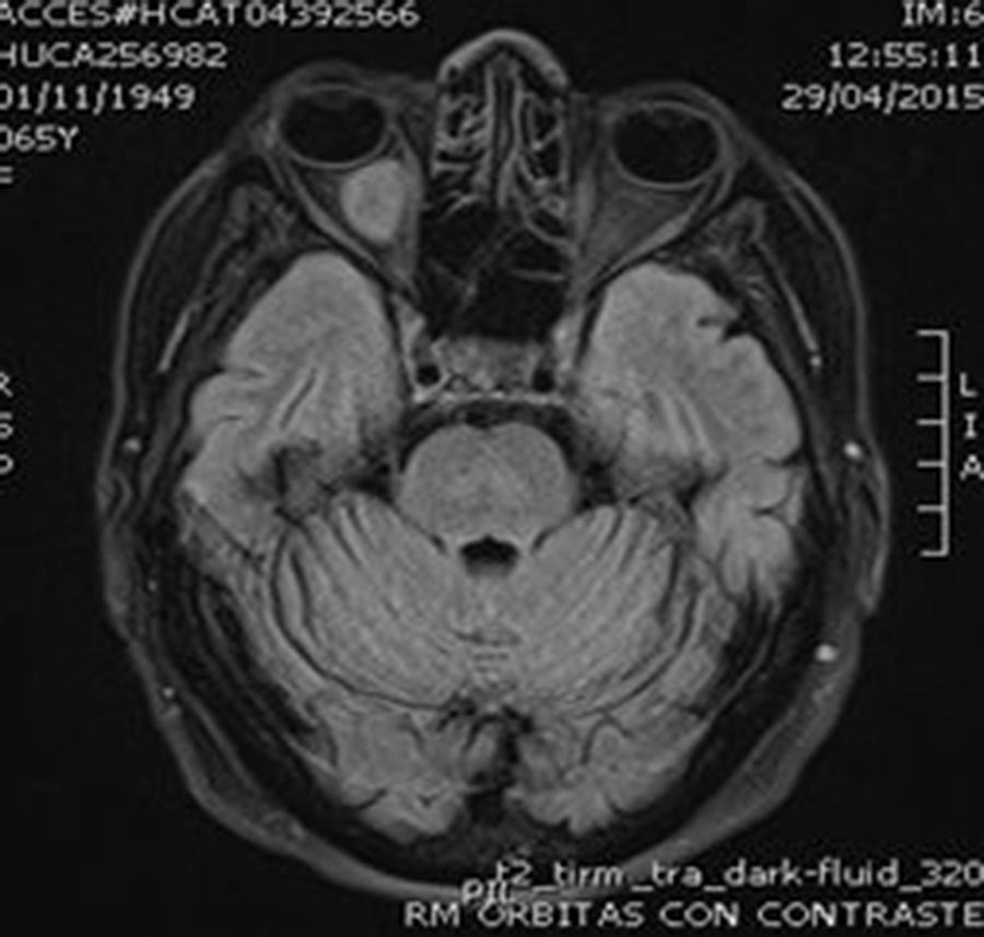
Our patient with sarcoidosis developed a retroocular mass, with a radiological differential diagnosis of sarcoidosis vs lymphoma. In sarcoidosis, thoracic lymph node involvement is characteristic and, although, it can also appear in other territories, it does so in fewer than 50% of the cases. As it is a systemic disease, in at least 10% of the patients it can also affect the central nervous system, generally, as leptomeningitis with infiltrations of the basal cisterns of the optic chiasm region and the hypothalamic-pituitary-adrenal axis. In these cases, it is essential to rule out infections and neoplasms, although magnetic resonance imaging or PET/CT confirm the diagnosis.2 In our patient, the diagnosis could be reached after biopsy of a supraclavicular lymph node. Despite presenting bone metabolic activity in PET/CT that suggested lymphomatous infiltration, it was found to be related to sarcoidosis.
The diagnosis of tumors in patients with sarcoidosis is well established, generally, after years of disease progress. However, the association with lymphoma referred to as sarcoidosis-lymphoma syndrome is more recent. In 1986, Brincker reported 17 cases of sarcoidosis that developed into Hodgkin's lymphoma.3 Since then, only a few cases have been described, only 3 in Spain.4–6 In contrast to the patients documented previously, those reported in Spain (Table 1) are characterized has being more elderly, and with a longer time to the diagnosis of the lymphoma.
Summary of the Spanish Patients With Sarcoidosis-lymphoma Syndrome.
| Gender | Clinical signs of sarcoidosis | Age at diagnosis of sarcoidosis | Clinical signs of progression | Type of lymphoma | Interval sarcoidosis-lymphoma | |
|---|---|---|---|---|---|---|
| 1 Hospital Universitari Germans Trias i Pujol (Barcelona) | Woman | Lymph node involvement, arthralgia, panniculitis and lung involvement | 66 years | Persistence of groin lymph node refractory to corticosteroids | NHL: DLBCL | ? |
| 2 Hospital Gregorio Marañón (Madrid) | Woman | Disseminated lymph node involvement, splenic infiltration and pulmonary nodules | 27 years | Neck discomfort with new lymph node changes | LH: predominantly pulmonary lymphocytic | 15 years |
| 3 Hospital La Paz (Madrid) | Woman | Peritoneal sarcoidosis | 64 years | Left hemithorax pain, discovery of infraclavicular mass | NHL: DLBCL | 3 years |
| 4 HUCA | Woman | Arthralgia and pulmonary involvement | 60 years | Diplopia and eye pain | NHL: DLBCL | 5 years |
DLBCL: diffuse large B-cell lymhoma; HL: Hodgkin's lymphoma; HUCA: Hospital Universitario Central de Asturias (a Spanish autonomous community [Principality of Asturias] in northern Spain); NHL: non-Hodgkin's lymphoma.
In short, we wish to bring light on the complexity of neurosarcoidosis, given the lack of specificity of the neuroimage and PET/CT, the difficulties for achieving a histological confirmation and the possibility of a lymphoma or infection. It is always necessary to insist on new biopsies, especially with lymph node involvement outside the clinical setting. In our patient population, individuals with sarcoidosis, are more likely to develop a non-Hodgkin's lymphoma and will, generally, be older than the usual mean age.
Ethical DisclosuresProtection of human and animal subjectsThe authors declare that no experiments were performed on humans or animals for this study.
Confidentiality of dataThe authors declare that no patient data appear in this article.
Right to privacy and informed consentThe authors declare that no patient data appear in this article.
Conflicts of InterestThe authors declare they have no conflicts of interest.
Please cite this article as: Brandy-García AM, Caminal-Montero L, Fernández-García MS, Saiz Ayala A, Cabezas-Rodríguez I, Morante-Bolado I. Síndrome sarcoidosis-linfoma. Reumatol Clin. 2016;12:339–341.


