Idiopathic hypoparathyroidism (IPTH) is a rare disorder that occurs in pediatric patients in the early years of life and is usually associated with autoimmune diseases.1 Hypoparathyroidism in adults is characterized by increased bone mineral density and ectopic calcification, but when it occurs in children before skeletal growth is complete it may also cause alterations in the rate of growth, bone remodeling and early epyphiseal closing.2
We present the case of a patient with a history of hypoparathyroidism from infancy that developed in the spine, with a “bone within bone” radiographic pattern in several vertebral bodies, which has been described infrequently in IPTH.
Clinical PresentationThe case is a 19-year-old woman, who was diagnosed with PIH at 10 years of age for limb dystonia secondary to hypocalcemia. She had no relevant personal or family medical history, including autoimmune and endocrine disorders. During follow-up she developed generalized bone sclerosis, which was evident both on radiographs of the pelvis and long bones of the lower extremities, and her bone densitometry showed lumbar spine (L1–L4) T-score values of 4.2, a Z-score of 4.5 and a BMD 1.513mg/cm2. She also developed bilateral cataracts which were operated on at the age of 12. Blood biochemistry showed mild hypocalcemia (calcium 7–8.5mg/dl), hyperphosphatemia (phosphorus above 5mg/dl), low levels of parathyroid hormone (4.8pg/ml) and normal alkaline phosphatase and vitamin D3 (25 [OH] D3 and 1.25 [OH] D3); she also showed increased urinary reabsorption of phosphate (95.3%) with normal calciuria. She received calcium supplements (calcium carbonate 2500mg/day), calcitriol (0.5g/day) and underwent a low phosphate diet.
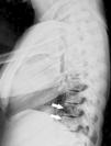
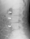
In imaging studies, in addition to marked bone sclerosis, her spine showed the presence of lines parallel to the cortex of the vertebral bodies giving rise to an image of a small copy of the vertebral body within the body, a sign called “bone within a bone” (Figs. 1–3). This sign was evident in several vertebral bodies. Morphometric vertebral deformities showed no calcification of paraspinal ligaments.
There are few publications on the presence of the “bone within a bone” sign in patients with IPTH. Rosen and Deshmukh described a series of 5 patients with IPTH, with this sign being the most common radiological finding, mostly affecting the lumbar vertebrae and in 2 cases with multiple affected vertebrae. This sign is related to the presence of lines parallel to the plates associated with the persistence of a remnant of the anterior vertebral surface. These lines may reflect the recovery of growth after a temporary cessation, for this reason are called “growth arrest recovery lines”. Additional factors that could explain the appearance of such lines in IPTH include treatment with high doses of vitamin D3 and changes in blood levels of calcium and phosphorus.2 There are other causes for the appearance of this “bone within bone” sign which can be classified according to the predominant mechanism: (a) new normal bone formation: normal neonates and infants, (b) abnormal periosteal new bone formation: prolonged administration of prostaglandin E1 in neonates, abnormal bone healing (not long after immobilized bone fractures, neuropathic disorders), infantile cortical hyperostosis (Caffey's disease), bone ossification of post-radiotherapy sarcoma, (c) splitting of the periosteal new cortical bone, secondary to bone infarcts (sickle cell and Gaucher's disease), scurvy, chronic osteomyelitis with entrapment and involucrum formation, (d) subcortical osteopenia, leukemia, metastasis, reflex sympathetic dystrophy, juvenile idiopathic osteoporosis, (e) alteration of the rate of bone growth: serious illness of acute or chronic diseases of intermittent course in pediatric patients that cause periods of increased bone resorption followed by inactive or quiescent periods, nutritional disorders (malnutrition, rickets), hypervitaminosis D, heavy metal poisoning, radiation, (f) failure or inhibition of osteoclast-mediated bone resorption: osteopetrosis, bisphosphonate treatment, (g) alteration of bone metabolism: Paget's disease, hereditary hiperphosphatasia (juvenile Paget's disease), acromegaly, (h) crystal deposition: oxalosis, and (i) iatrogenic or false findings: bone grafts, subcortical bone cement, spiral fracture, an incidental finding during normal consolidation of a fracture and artifacts due to technical factors.3
Ethical disclosuresProtection of human and animal subjects. The authors declare that no experiments were performed on humans or animals for this investigation.
Confidentiality of Data. The authors declare that they have followed the protocols of their work centre on the publication of patient data and that all the patients included in the study have received sufficient information and have given their informed consent in writing to participate in that study.
Right to privacy and informed consent. The authors declare that no patient data appears in this article.
Please cite this article as: Sifuentes Giraldo WA, et al. El signo del «hueso dentro de hueso» en el hipoparatiroidismo idiopático. Reumatol Clin. 2012;8:372–4.


