


To determine the prevalence of vitamin D deficiency in patients with small and medium vessel systemic vasculitis.
MethodsIn this cross-sectional study, 25-hydroxy (OH) vitamin D3 levels were measured in adult patients with systemic small and medium vessel vasculitis including antineutrophil cytoplasmic antibody-associated vasculitis (AAV), cryoglobulinaemic vasculitis (CryV), IgA vasculitis (IgAV) and polyarteritis nodosa (PAN), and age- and sex-matched healthy subjects (HS) and patients with rheumatoid arthritis (RA) as control groups. 25OH vitamin D3 levels<30ng/ml and <20ng/ml were regarded as insufficiency and deficiency, respectively.
ResultsFifty-seven patients (42 AAV, 2 CryV, 8 IgA vasculitis, 5 PAN) with systemic vasculitis, 101 HS, and 111 RA patients were included. The mean 25OH vitamin D3 level was 21.8±14.2ng/mL in patients with vasculitis, 42.7±27.6ng/mL in HS (p<.001) and 20.1±18.47ng/mL in patients with RA (p=.54). Vitamin D insufficiency and deficiency were significantly higher in patients with systemic vasculitis compared to HS (75.4% vs 33.7%, p<.001; %50 vs 21.8%, p<.001, respectively). Vitamin D status was not different in patients with systemic vasculitis compared to RA. There was a negative correlation between vitamin D status and CRP levels (=−.364, p=.007). The multivariate logistic regression analysis showed that renal involvement was significantly associated with vitamin D deficiency/insufficiency in patients with vasculitis (OR 22.5 [95% CI 1.6–128.9].
ConclusionVitamin D deficiency and insufficiency are more frequent in patients with systemic small and medium vessel vasculitis and RA than HS. Renal involvement is one of the factors associated with vitamin D deficiency/insufficiency in patients with vasculitis.
Determinar la prevalencia de la deficiencia de vitamina D en los pacientes con vasculitis sistémica de pequeños y medianos vasos.
MétodosEn este estudio transversal se midieron los niveles de 25-hidroxivitamina D3 en pacientes adultos con vasculitis sistémica de pequeños y medianos vasos, incluyendo vasculitis asociada a anticuerpos anticitoplasma de neutrófilos (AAV), vasculitis crioglobulinémica (CryV), vasculitis IgA (IgAV) y poliarteritis nodosa (PAN), y sujetos sanos pareados por edad y sexo (SS) y pacientes con artritis reumatoide (AR) como grupos control. Se consideraron insuficientes y deficientes los niveles de 25-hidroxivitamina D3<30ng/ml y <20ng/ml, respectivamente.
ResultadosSe incluyeron 57 pacientes (42 de AAV, 2 de CryV, 8 de vasculitis IgA y 5 de PAN) con vasculitis sistémica, 101 SS y 111 pacientes de AR. El nivel medio de 25-hidroxivitamina D3 fue de 21,8±14,2ng/ml en pacientes con vasculitis, 42,7±27,6ng/ml en SS (p<0,001) y 20,1±18,47ng/ml en pacientes con AR (p=0,54). La insuficiencia y deficiencia de vitamina D fueron significativamente más altas en los pacientes con vasculitis sistémica en comparación con los SS (75,4 vs. 33,7%; p<0,001; 50 vs. 21,8%; p<0,001, respectivamente). El estatus de vitamina D no fue diferente en los pacientes con vasculitis sistémica en comparación con AR. Existió una correlación negativa entre el estatus de vitamina D y los niveles de PCR=−0,364; p=0,007. El análisis de regresión logística multivariante reflejó que el compromiso renal estuvo significativamente asociado a la deficiencia/insuficiencia de vitamina D en los pacientes con vasculitis (OR: 22,5; IC 95%: 1,6-128,9).
ConclusiónLa insuficiencia y deficiencia de vitamina D son más frecuentes en los pacientes con vasculitis sistémica de pequeños y medianos vasos y AR que en los SS. El compromiso renal es uno de los factores asociados a la deficiencia/insuficiencia de vitamina D en los pacientes con vasculitis.
Vitamin D is a fat-soluble vitamin that has a significant clinical role in calcium hemostasis and bone metabolism. Besides, it has been shown that vitamin D has intracellular receptors in the endothelium, smooth muscle, myocardium, brain, prostate, breast, intestine, and immune cells.1 Therefore, vitamin D and its metabolites are shown to mediate important regulatory effects in the body such as anti-hypertensive, anti-atherosclerotic and anti-inflammatory on adaptive immune response and antimicrobial response on innate immune response associated with vitamin D receptors and other mechanisms.2–7 Vitamin D receptor is present also on antigen-presenting cells, macrophages, B, CD4+ and CD8+ T lymphocytes, and plays a role in autoimmune diseases by providing T cell tolerance to self-antigen.8,9 In this regard, vitamin D levels have been studied in various autoimmune rheumatic diseases such as rheumatoid arthritis (RA), systemic lupus erythematosus (SLE), Behcet's disease and Takayasu's arteritis.6,8,10–15
It has been shown that vitamin D levels were lower in patients RA who had high disease activity compared to patients with the inactive disease.10 A significant inverse correlation between serum vitamin D levels and RA disease activity was also shown.11 Moreover, remission rates and response to treatment were significantly lower in RA patients with vitamin D deficiency than those with normal vitamin D levels.10–13 It has also known how vitamin D regulates the pathologic mechanism of RA.16 Similarly, SLE patients were found to have lower vitamin D levels compared to healthy controls and vitamin D levels inversely correlated with SLEDAI.6,8,14,15
Systemic vasculitides are chronic inflammatory disorders with significant morbidity and mortality and share similar immunopathological mechanism with RA.17,18 Vitamin D can affect the course of vasculitis disease with the effects on the immune system. Despite the existent evidence of the immunomodulatory effects of vitamin D in other autoimmune diseases, there are only a few studies of vitamin D in systemic vasculitis limited to ANCA-associated vasculitis(AAV), Takayasu's arteritis(TAK) and Behcet's Disease(BD).19,20
It is important to know the vitamin D status in patients with systemic vasculitis, both to prevent potential complications of long-term glucocorticoid use and improve the immune functions that can alter disease severity. In this study, we sought to assess the frequency of vitamin D deficiency in patients with small and medium vessel vasculitis in comparison to healthy subjects (HS) and patients with RA.
Patients and methodsPatients with systemic vasculitides including antineutrophil cytoplasmic antibody (ANCA)-associated vasculitis, cryoglobulinemic vasculitis(CryV), IgA vasculitis(IgAV) and polyarteritis nodosa(PAN) were recruited from Marmara University, School of Medicine Hospital Rheumatology and Nephrology Clinics between January-March 2015 and October-March 2016. All patients fulfilled ACR classification criteria for PAN, cryoglobulinemic vasculitis and IgA-vasculitis, and the 2012 Chapel Hill Consensus definitions for ANCA-related vasculitis.17,21 Age- and sex-matched healthy subjects (HS) without a chronic inflammatory disease from Internal Medicine clinic who were followed for routine healthcare checks and as another disease-control group, sex- and age-matched patients with RA from the same Rheumatology clinic were also recruited. All RA patients fulfilled the 2010 ACR/EULAR classification criteria for RA. Patients and HS who were already on any type of vitamin D replacement or pregnant or breastfeeding had active cancer or infection, parathyroid disease, or calcium abnormalities were excluded from the study.
Data on sociodemographics, disease duration, disease activities, and all medications taken were determined on the day of recruitment through patient charts. The study was approved by the Marmara University Local Ethics Committee (Ethics Committee protocol code: 09.2014.0227). Informed consent was obtained from all patients and HS participating in the study according to the Declaration of Helsinki.
Serum vitamin D level measurementsVitamin D levels may vary depending on the season and the latitude of the region. Therefore, all samples were collected during January–March and October–March period. Serum vitamin D was determined as 25-hydroxy (OH) vitamin D3 levels by using Roche Elecsys and Cobas immunoassay method which uses the electrochemiluminescence immunoassay ECLIA method. All vitamin D values used in this study indicate values of serum 25OH vitamin D3 levels.
Vitamin D levels of ≥ 30 ng/ml were considered as adequate, <30ng/ml and >20ng/ml as insufficient, and levels≤20ng/ml as deficient. Patients and HS with vitamin D insufficiency and deficiency were recommended to use 50,000IU cholecalciferol weekly for the first eight weeks and 10 drops (1330IU/day) for at least 6 months. At the end of a one-year follow-up, serum vitamin D levels were measured again at the same seasonal period with the same technique.
Statistical analysisThe conformity of the variables to normal distribution was examined by visual (histogram and probability graphs) and analytical methods (Kolmogorov–Smirnov/Shapiro–Wilk tests). Descriptive analyzes were performed using the mean±standard deviation (SD) for the normally distributed variables, and median and interquartile range (IQR) for variables not normally distributed. Demographic characteristics and vitamin D levels of study and control groups were compared by the chi-square and Student t-test or Mann–Whitney U test. Correlation between disease characteristics and clinical parameters and levels of vitamin D were assessed by Pearson or Spearman correlation test according to data distribution. A stepwise multivariate logistic regression analysis was performed to determine the disease characteristics that may be associated with vitamin D deficiency/insufficiency in patients with vasculitis. The following covariables were analyzed in the multivariate model: gender, age, vasculitis duration, renal, gastrointestinal and cardiovascular involvement, c-reactive protein (CRP), the daily dose of prednisone, type of vasculitis. Hosmer–Lemeshow goodness of fit was used to evaluate the model fit. Statistical analyses were performed using the Statistical Package for Social Sciences (SPSS for Windows, 16.0, IL). Statistical significance was indicated by p values less than 0.05.
ResultsA total of 57 vasculitis patients (F/M=22/35, mean age: 51.0±16.4 years, median 22[IQR] se duration of 3 [1–7] years), 101 HS (F/M=62/49, mean age:48.6±14.2 years) and 111 RA patients (F/M=59/52, mean age: 52.9±11.77, median [IQR] disease duration 11 [8-16] years) were studied (Table 1). In the vasculitic patient group, 42 had AAV(granulomatous polyangiitis: 28, microscopic polyangiitis[MPA]:14), 2 had CryV [both negative for hepatitis C], 8 had IgAV and 5 had PAN. Thirty-nine (68.4%) patients were on glucocorticoid (GC) treatment at the time of recruitment. Patients had different treatment modalities at different time of their illness. Of patients 36(63.2%) had on cyclophosphamide, 36(63.2%) azathioprine, 17(29.8%) rituximab, 12(21.1%) plasmapheresis, 5(8.8%) methotrexate, and 2(3.5%) mycophenolate mofetil treatment.
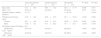
Demographic characteristics of patients with vasculitis and healthy individuals and 25 hydroxy vitamin D3 levels.b
| Vasculitis patientsa (n=57) | Healthy subjects (n=101) | RA patients (n=111) | P1 value | P2 value | |
|---|---|---|---|---|---|
| Age, years | 51.0±16.4 | 48.6±14.2 | 52.9±11.77 | 0.328 | 0.431 |
| Male, n (%) | 35 (61.4) | 49 (48.5) | 52 (46.8) | 0.127 | 0.074 |
| Disease duration, median (IQR) yrs | 3 (1–7) | – | 11 (8–16) | – | <0.001 |
| 25hydroxyvit-D3 level, ng/ml | 21.8±14.2 | 42.6±27.6 | 20.1±18.47 | <0.001 | 0.539 |
| Female | 24.7±16.8 | 37.9±27.4 | 20.92±22.76 | 0.038 | 0.478 |
| Male | 19.9±12.1 | 51.1±26.2 | 19.1±12.0 | <0.001 | 0.755 |
| 25 hydroxy vitamin D3 status, n (%) | |||||
| Insufficiency (<30ng/ml) | 43 (75.4) | 34 (33.7) | 92 (82.9) | <0.001 | 0.252 |
| Deficiency (<20ng/ml) | 29 (50.9) | 22 (21.8) | 66 (59.5) | <0.001 | 0.289 |
The baseline serum vitamin D levels of the patients with vasculitis were significantly lower than the HS (21.8±14.2ng/ml vs. 42.6±27.6ng/ml, p<0.001) (Table 1). Forty-three of the 57 vasculitis patients (75.4%) had vitamin D deficiency and insufficiency. Deficiency and insufficiency of vitamin D were significantly higher in patients with vasculitis compared to HS (50.9% vs 21.8% for deficiency and 75.4% vs 33.7% for insufficiency). However, deficiency and insufficiency of vitamin D were similar in patients with vasculitis compared to RA patients (50.9% vs 50.9% for deficiency and 75.4% vs 82.9% for insufficiency) (Table 1). Both male and female patients with vasculitis had similar levels of vitamin D, however, male HS had higher vitamin D levels than female HS (Table 1). However, vitamin D levels of female and male patients with vasculitis were significantly lower than those of the corresponding HS (Table 1).
There were no significant differences in vitamin D levels according to the vasculitis type. We have compared the vitamin D levels of patients with AAV and other vasculitis, and there was no difference. There was no correlation between vitamin D levels and ANCA positivity. Deficiency and insufficiency of vitamin D were similar in patients with AAV vasculitis compared to non-AAV vasculitis patients (46.5% vs 64.3% for deficiency and 74.4% vs 78.6% for insufficiency, p=0.25 and 0.75, respectively).
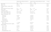
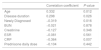
When the factors associated with vitamin D deficiency/insufficiency were investigated in vasculitis patients, we observed that vasculitis patients with vitamin D deficiency/insufficiency were younger than those with sufficient vitamin D, but there was no difference according to the duration of the disease and GC use (Table 2). Vitamin D levels also positively correlated with age and vasculitis duration and negatively correlated with CRP levels and a new diagnosis of vasculitis disease (Table 3.). The multivariate logistic regression analysis showed that renal involvement was significantly associated with vitamin D deficiency/insufficiency (OR 22.5[95% CI 1.6–128.9]).
Clinical features of vasculitis patients with and without 25 hydroxy vitamin D3 deficiency.a
| Patients with sufficient vitamin D (n=14) | Patients with insufficient/deficient vitamin D (n=43) | P value | |
|---|---|---|---|
| Age, years | 60.6±9.5 | 47.9±16.9 | 0.011 |
| Male, n (%) | 5 (35.7) | 30 (69.8) | 0.023 |
| Disease duration, median (IQR) years | 3 (0–18) | 2.5 (1–6) | 0.391 |
| Daily prednisolone dose, mg/day | 54±35 | 66±26 | 0.154 |
| BUN, mg/dL | 23.28±9.1 | 26.87±19.21 | 0.35 |
| Creatinine, mg/dL | 1.2±0.44 | 2.1±3.0 | 0.07 |
| ESR, median (IQR) mm/h | 35 (21–52) | 30 (18–51) | 0.379 |
| CRP, median (IQR) mg/dL | 3.4 (1.3–6.2) | 3.4 (3.2–12.5) | 0.196 |
| Organ involvement, n (%) | |||
| Cutaneous | 5 (35.7) | 19 (44.2) | 0.577 |
| Pulmonary | 7 (50) | 23 (53.5) | 0.820 |
| Renal | 11 (78.6) | 32 (74.4) | 0.754 |
| Neurological | 6 (42.9) | 10 (23.3) | 0.156 |
| Cardiovascular | 0 | 3 (7) | 0.310 |
| Vasculitis subclass | |||
| AAV, n (%) | 11 (78.6) | 32 (74.4) | 0.75 |
| Daily prednisolone dose, mg/day, Median (IQR) | 4 (6.5) | 4 (4.0) | 0.852 |
| CYC, n, (%)b | 9 (64.3) | 27 (62.8) | 0.920 |
| AZA n, (%)b | 8 (57.1) | 28 (65.1) | 0.591 |
| RTX n, (%)b | 1 (7.1) | 16 (37.2) | 0.033 |
| MTX n, (%)b | 0 (0) | 5 (11.6) | 0.182 |
| Plasmapheresis n, (%)b | 2 (14.3) | 10 (23.3) | 0.475 |
Correlation of vitamin D3 levels with demographic characteristics and disease activity parameters of patients.
| Correlation coefficient | P value | |
|---|---|---|
| Age | 0.332 | 0.012 |
| Disease duration | 0.298 | 0.026 |
| Newly Diagnosed | −0.319 | 0.016 |
| BUN | −0.021 | 0.878 |
| Creatinine | −0.127 | 0.346 |
| ESR | −0.081 | 0.561 |
| CRP | −0.364 | 0.007 |
| Prednisone daily dose | −0.104 | 0.442 |
ESR, erythrocyte sedimentation rate; CRP, C-reactive protein; BUN, blood urea nitrogen.
Vitamin D levels of 33 vasculitis patients who were vitamin D deficient or insufficient were re-evaluated 1 year after the first evaluation. The baseline vitamin D level of these 33 patients was 22.5±14.8ng/ml and increased to 26.6±14.2ng/ml in the first year without a significant difference (p=0.18). At 1 year, 26(78.8%) of 33 patients who had baseline vitamin D deficiency or insufficiency were still vitamin D insufficient (<30ng/ml). Although cholecalciferol treatment was recommended to all vitamin D deficient/insufficient vasculitis patients, only 17(51.5%) of those were adherent to the vitamin D replacement (Table 4).
Comparison of vasculitis patients with and without vitamin D replacement.
| Receive cholecalciferol(n=17) | Did not receive cholecalciferol(n=16) | P value | |
|---|---|---|---|
| Age, years | 52.4±14.1 | 46.9±15.7 | 0.390 |
| Male, n (%) | 12 (75) | 11 (64) | 0.052 |
| 25 Hydroxy vitamin D3 (ng/ml) | 31.9±17.1 | 20.9±7.4 | 0.024 |
| 25 Hydroxy vitamin D3 status, n (%) | |||
| Sufficient | 9 (52.9) | 3 (18.8) | 0.090 |
| Insufficient/deficient | 8 (47.1) | 13 (81.2) | 0.044 |
Vitamin D levels of those 17 patients who received cholecalciferol increased from 19.9±10.9ng/ml to 31.8±17.1ng/ml (p=0.006). Among those received replacement treatment, 9(%52.9) achieved sufficient vitamin D levels.
DiscussionVitamin D has shown to have immunomodulatory and immunosuppressive properties as a steroid hormone besides its well-known effects on bone and calcium metabolism. Thus, it is important to know the vitamin D levels in systemic inflammatory rheumatic diseases where immunomodulators that can also adversely influence bone health are also frequently used. In this study, we observed that patients with systemic small/medium vessel vasculitis had significantly more frequent vitamin D deficiency/insufficiency compared to healthy subjects. However, their vitamin D levels were similar to age- and sex-matched RA patients. The most important factors are sun exposure which is free and available for all people and the dietary sources of vitamin D. So for any population sharing more or less similar social standards, there will be no major discrepancies between vitamin D levels associated with these factors. We think that the significant difference between the vasculitis group and those normal healthy subjects arise from the disease itself or the factors with which it is associated. The main determinant of vitamin D deficiency/insufficiency in vasculitis patients was renal involvement. Although all vitamin D deficient/insufficient vasculitis patients were recommended vitamin D replacement, only half was compliant on this regimen.
Although there was a negative correlation between BUN and creatinine levels with Vitamin D levels in correlation analysis, it was not statistically significant. However, the multivariate logistic regression analysis showed that renal involvement was significantly associated with vitamin D deficiency/insufficiency (OR 22.5 [95% CI 1.6–128.9]). It is well-known that patients with renal disease can have inadequate vitamin D levels. Therefore, we added renal disease as a covariate in the logistic regression analysis with an outcome of “Vitamin D deficiency/insufficiency.” This variable was a binary variable (Vit D<30 or ≥30) rather than the continuous variable used in the correlation analysis. Furthermore, although some patients had renal involvement in our study, not all had a persistently elevated creatinine (improved with treatment) as we included both newly diagnosed and established patients. We believe that the reason we did not find a significant correlation with creatinine but a significant association with kidney disease is related to this.
Although, vitamin D levels have been studied in RA and SLE, there have been few studies assessing the relationship between systemic vasculitis and vitamin D in the literature, mainly Takayasu's arteritis and Behcet's Disease.20 Favorable effects of vitamin D treatment on endothelial function are reported.19,20 In a recent study conducted in patients with TAK and BD, vitamin D level was 16.9±10.6nmol/L in TAK and 38.8±20.9nmol/L in BD patients. The frequency of vitamin D deficiency was 83.3%, 46.5%, and 3.3% in patients with TAK, BD, and healthy controls, respectively.20 Vitamin D level was lower in active BD patients compared to inactive ones and there was a positive correlation between vitamin D level and T regulatory lymphocyte number in another study.23In the general population, the most well-known risk factor for vitamin D deficiency is inadequate sunlight exposure. In studies evaluating the relationship between frequency of AAV and UV radiation level, it was found that the incidence of GPA and eosinophilic granulomatosis with polyangiitis increased with increasing latitudes and decreasing UV radiation levels. This association was thought to be due to the decreased vitamin D levels in people living high latitudes.24
In a study investigating the relationship between HCV-associated cryoglobulinemia and vitamin D, it was shown to be associated with the presence of mixed cryoglobulinemia with vitamin D<13ng/ml.25 In another study Cusato et al. showed that vitamin D receptor polymorphism can be associated with mixed cryoglobulinemia presence in HCV-infected patients.26 Another study also showed the association between serum 25OH vitamin D3 levels and the presence of HCV-related MC and CryV.27
In a study evaluating the risk factors for relapses after ANCA positivity, relapse was detected in 36 of 60 patients with ANCA titer increase, especially in the autumn season. In patients with ANCA titer increase, vitamin D levels decreased significantly in patients with a relapse and slightly increased in remission (difference −6.3±14.4, p=0.017 versus 2.7±16.3, p=0.430).28 In this study, it was suggested that in addition to an increase in ANCA titers, a second factor that may cause an increase in inflammation may be required for the development of relapse and one of these factors may be vitamin D.28
In studies examining vitamin D levels in RA patients, vitamin D deficiency and insufficiency were found to range between 29–72%% and 64.5–90.9% which was similar to our rates for RA.29 Similarly, a study examining vitamin D levels in the Turkish general population reported that vitamin D deficiency and insufficiency in Turkish people were 72% and 90.9%, similar to our rate.30
Our study is the first study to evaluate vitamin D levels in patients with small/medium vessel vasculitis. Besides finding frequent vitamin D deficiency/insufficiency in patients with vasculitis, we also observed that vitamin D deficiency/insufficiency was more common in young patients. The reason for this may be the more frequent use of over the counter vitamin D supplementations by older patients. Vitamin D deficiency is common in patients with chronic kidney disease, particularly in patients with proteinuria, due to loss of 25-hydroxyvitamin D with vitamin D binding protein.31 In this study, we also found that vasculitis patients with renal involvement had a higher risk of being vitamin D deficient/insufficient.
Another noteworthy finding of our study was the low adherence rates to vitamin D replacement treatment. Among the treatment recommended patients, only half used the recommended vitamin D treatment, and only half of those received vitamin D achieved sufficient vitamin D levels. Intake of vitamin D supplements in particular in individuals with low-socioeconomic status is limited by relatively poor adherence.32 Given the complexity of the systemic vasculitis and several immunomodulatory medications started for management, polypharmacy may be a reason for this low adherence rates in vasculitis patients. As vitamin D have both immunomodulatory and bone-related positive effects in these patients on glucocorticoids, higher doses of vitamin D may be considered for replacement. Physicians, therefore, should question compliance during the follow-up visits in vasculitis patients.
Our study has some limitations. As a cross-sectional study, we could not assess the influence of vitamin D on disease activity on a stable immunosuppressive regimen in patients during remission. As both newly diagnosed and established vasculitis patients were included, vitamin D levels can be influenced by different immunomodulatory medications, hospitalizations, or significant damage caused by vasculitis. However, we tried to collect all samples at the same season to, at least, minimize one of the most important factors determining vitamin D levels, sunlight exposure. Another limitation was the low adherence of patients to vitamin D treatment. Compliance of patients to treatment and recommended dosage should be followed at regular intervals.
As a conclusion, vitamin D deficiency/insufficiency is quite common in patients with systemic small/medium size vasculitis. Renal involvement and avoiding the sun and lack of nutrition that are precipitated by a significant systemic disease may be the contributing factors to this deficiency. Vitamin D adherence rates were also low in our patient group. As vitamin D has well-known immunomodulatory properties, all vasculitis patients should be screened and treated for vitamin D deficiency. They should also be monitored for adherence to the treatment of vitamin D deficiency. Moreover, a longer and higher dose vitamin D supplementation can also be considered in patients with vasculitis patients.
Conflicts of interestNone to declare.
22.


