

Currently, the measurement of bone remodeling biomarkers is an innovate proposal in clinical evaluation of patients with osteoporosis. Its use may identify patients at an increased risk of fracture as well as monitoring therapeutic efficacy. Because they constitute a relatively inexpensive non-invasive measurement, its use should be widespread for serial and frequent measurements of bone turnover. However, their analytical and biological variabilities limit their clinical applicability.
En la actualidad, la determinación de marcadores bioquímicos de remodelado óseo supone una propuesta novedosa en la evaluación clínica de los pacientes con osteoporosis. El uso de estos biomarcadores podría permitir la identificación de pacientes con mayor riesgo de fractura y monitorizar la respuesta terapéutica. Al tratarse de mediciones no invasivas y relativamente económicas, debería extenderse su empleo, ya que posibilitaría una medición seriada y en intervalos cortos de las variaciones en el recambio óseo. Sin embargo, su variabilidad analítica y biológica limita en la actualidad su aplicabilidad clínica.
In adults, bone tissue undergoes a constant process of replacement involving the osteoclast, leading to bone resorption, and osteoblasts, responsible for the compensatory phase of bone formation. The bone remodeling cycle is completed in a period of 3–6 months, predominating the formative phase over resorption. Under physiological conditions there is a balance between the two. However, when a decoupling occurs due to a preponderance of resorption, bone mass loss occurs, commonly leading to osteoporosis.1
Quantitative measurements of bone mineral density (BMD) are essential for the clinical evaluation of patients with osteoporosis but, being a static parameter, provides no information of the rate of bone turnover. In contrast, biochemical markers of bone remodeling offer a dynamic, global analysis of the skeleton.2
Biochemical Markers of Bone RemodelingThe organic matrix of bone is 90% constituted by type I collagen. During the degradation process, there is extracellular release of the carboxy peptides and amino protocollagen molecules, which then pass into the blood stream.3 Biochemical markers of bone turnover measure these products generated during the process of formation or degradation of bone matrix and can be determined in blood and urine. Their analysis, repeated at short intervals, allows a serial assessment of bone turnover. Measuring bone markers of osteoblastic activity are referred to as formation markers and those derived from the activity of osteoclasts are called resorption4 markers (Table 1).
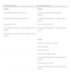
Markers of Bone Remodeling.
| Formation Markers | Resorption Markers |
| Serum | Serum |
| Total alkaline phosphatase (FA) | Tartrate resistant acid phosphatase (TRAP) |
| Bone alkaline phosphatase (FAO) | C-terminal telopeptide of collagen type I (ICTP) |
| Osteocalcin (OC) | β-CrossLaps (β-CTX) |
| C-terminal propeptide of protocollagen type I (PICP) | N-terminal telopeptide of collagen type I (NTX) |
| N-terminal propeptide of protocollagen type I (PINP) | |
| Urine | |
| Urinary excretion of calcium | |
| Hidroxyproline | |
| Pirydinolin (Pir) | |
| Deoxypirydinolina (Dpir) | |
| C-terminal telopeptide of collagen type I (ICTP) | |
| α-CrossLaps (α-CTX) | |
| N-terminal telopeptide of collagen type I (NTX) |
Adapted from: Torres et al. Endocrinol Nutr 2003;50(6):72.
For the measurement of bone-forming activity there are several clinically useful markers: alkaline phosphatase, osteocalcin and protocollagen I extension peptides.
Alkaline phosphatase activity is derived from various tissues such as the liver, bone, placenta, etc. Bone and liver isoforms are the most common (90%). Both are found in the same proportion in the healthy individual and differ in glycosylation patterns, and there is a cross-activity of 10%–20%, according to studies with monoclonal antibodies.5 The bone isoform has the advantage of presenting no variation between genders and not influenced by circadian rhythm, so that, despite having low sensitivity and specificity in the study of metabolic bone disease, it is easy to detect in the absence gestation and liver disease.6
Osteocalcin is the most abundant non-collagenous protein of the extracellular matrix. Specific for bone and dentin, it is elevated in situations of increased bone turnover, has a short half-life and is eliminated via the urine, so its levels are increased in situations of renal failure.7 Its exact role in bone remodeling is not well established. Recent work has analyzed the potential role of infracarboxilated osteocalcin in predicting bone mass and risk of fracture.8
Because type I collagen is the main product of synthesis of the osteoblast, the amino-terminal carboxy propeptides would, theoretically, be the ideal marker of bone formation. However, the fact that type I collagen appears in other tissues other than bone limits its use in the study of metabolic bone disease.9
Resorption MarkersHistorically, urinary calcium was the first test used to assess bone resorption. However, the fact that it is influenced by various factors, such as calcium intake, intestinal absorption and renal threshold of excretion of calcium, makes its determination a test with low sensitivity and specificity, and is currently unused. The same applies to urinary hydroxyproline: different tissue origins and the influence dietary intake exerts on it are to be considered in its low yield.1
The collagen molecules in bone matrix are covalently linked by pyridinoline (Pir) and deoxypyridinoline (Dpir) forming fibrils. The former is also found in cartilage, however, Dpir is more specific for bone.10 They do not depend on diet, and are not absorbed via the gut. They express both changes of bone metabolism, rising in childhood, menopause, osteomalacia, hyperparathyroidism and hyperthyroidism, and lowering due to estrogen and bisphosphonate treatment.11
Other elements released during bone resorption are the carboxyterminal (ICTP, CTX) and amino-terminal (NTX) telopeptides of collagen. They have shown a significant correlation with BMD in postmenopausal women, and both CTX and NTX are considered the most clinically useful markers of bone resorption currently available.12,13
Tartrate-resistant acid phosphatase 5b (5b Fatra) is a lysosomal enzyme not only involved in osteoclast bone degradation, but is also present in other tissues. It is poorly specific, and together with the methodological difficulty in identifying it, currently makes it of little use.14
Other Biochemical Markers of Bone RemodelingThere are newly developed bone turnover markers whose clinical value is still under study. Cathepsin K acts to degrade the collagen and its serum levels reflect the number of osteoclasts; it has been suggested as a marker of bone resorption. Urinary osteocalcin fragments also come from the resorption of bone matrix and appear to be more specific. In the assessment of bone quality posttranslational modifications of bone matrix proteins such as infracarboxylated osteocalcin, native and isomerized forms of CTX and pentosydine have been considered especially important.13,15
Clinical Utility of Biomarkers in OsteoporosisThe most important current clinical application of markers of bone turnover in osteoporosis is the assessment of therapeutic response. They have also been useful in predicting risk of fracture and bone loss and their correlation with BMD. However, the results of these have been mixed depending on the type of study population and marker analyzed.16 Regarding the prediction of bone mass, although biomarkers evaluate the balance between bone formation and resorption and are generally inversely related to BMD, these correlations are not strong enough to predict bone mass, therefore, should not be used for diagnosing osteoporosis.17
Control of the Therapeutic EfficacyCurrently represents its best established clinical use. Several studies have shown that after initiating antiresorptive therapy there is a significant decrease in both resorption (within 4–6 weeks), and in bone formation markers (2–3 months). In most cases there is a low value reached 2–3 months after start of treatment and remains constant while the patient continues taking the drug. A significant change would be between 40% and 70% of reduction in markers of bone resorption (CTX in serum and urine NTX and Dpir in urine), when using a potent antiresorptive (bisphosphonates), and more modest reductions (30%–40%) with less energetic catabolic (raloxifene). Therefore, the changes will depend on the therapeutic agent employed and the marker analyzed.
Thus, the failure to observe these reductions indicates poor adherence to treatment by the patient or the improper administration of the drug.18–20
The bone forming treatment initially induces a rapid increase in bone formation markers, followed by a subsequent increase in resorption markers. In this regard, several studies have pointed to markers such as PICP and especially PINP as they have a higher sensitivity to predict changes in BMD in patients treated with teriparatide. A proposed performance algorithm in patients treated with this anabolic agent is related to the variation detected in PINP: increases above 10μg in this formation marker, 3 months after initiation of therapy, suggest an appropriate response to it.21,22
Although it is desirable to have markers of formation and resorption, according to the available evidence the markers of bone turnover that are more sensitive and clinically useful are serum CTX—when using an antiresorptive—and PINP when treating with anabolic drugs. Their determination, after 2 or 3 months of treatment, offers the remarkable advantage of being able to assess the effectiveness of medication and reassuring patients without having to wait 12–24 months to document changes in BMD.23
Predicting Response to TreatmentSeveral studies have demonstrated an association between changes in bone turnover markers after antiresorptive or anabolic treatment and long-term anti-fracture efficacy. A meta-analysis of 18 clinical trials found that, during antiresorptive therapy, a 70% decrease in resorption markers decreased the risk of vertebral fracture by 40% and a decrease of 50% of formation markers reduced this risk by 44%.24 In this sense, there is evidence that an increase in formation markers, such as PIPC and FAO, a month of starting treatment with teriparatide was associated with improvement in bone structure.25
However, the optimal threshold for each marker is not well established and there is no long-term data when the biomarkers are reduced below the reference range. Some researchers support the hypothesis that it could interfere with physiological bone repair of “microcracks” and, therefore, lead to increased bone fragility.26
Selection of Patients to TreatIt has been suggested that patients with a greater increase in bone turnover markers respond better to antiresorptive therapy. In a 2-year prospective study in women with hormone replacement therapy, patients placed in higher quartiles for urinary excretion of NTX showed a higher bone gain.27
Theoretically, the reference levels of biomarkers could be used to guide treatment decisions more rationally. However, most clinical trials have shown that anti-fracture efficacy is largely independent of the baseline markers of bone remodeling. Therefore, it is not considered currently as clinically useful.28
Prediction of Fracture RiskBMD measured by DXA remains the best predictor of fracture risk. However, BMD does not explain everything and must take into account other variables, including the rate of bone turnover. Data from several prospective studies in elderly postmenopausal women show an association, independent of BMD, between the indices of bone turnover and osteoporotic fracture risk, although the results are discordant, particularly with regard to bone29 formation markers. Elevated bone resorption markers have been associated with an increase of approximately twice the risk of vertebral and nonvertebral fractures compared with women in whom such markers were at normal levels. The EPIDOS study results were consistent with this fact: In older women, increased bone resorption markers in urine (CTX) were associated, after adjustment for BMD, with an increased risk of hip fracture. This suggests that a combined approach, using BMD and biochemical markers of bone remodeling, could improve the prediction of fracture risk in postmenopausal women, and this risk increases in those with low BMD and/or elevated markers of bone remodeling.30
Prediction of Bone LossAnother prominent use of these biomarkers is the possibility of identifying women who will present a high rate of bone loss in the years following menopause, in order to start a preventive strategy for osteoporosis. As is known, bone densitometry is an important predictor of fracture risk, but a single measurement does not portend the rate of bone loss. Several studies have shown that markers of bone turnover may be useful in determining the rate of bone mass loss,1 with markers of resorption providing greater predictive power than those of formation.5,31
A recent review has analyzed the association of biomarkers with loss of bone mass in different skeletal sites. The most consistent relationship has been detected at the distal radius and calcaneus. In the hip, the relationship is more modest, while in the lumbar spine it is negligible. The latter finding could be due to the high prevalence of vertebral osteoarthritis in the elderly. In summary, markers of bone turnover together with other demographic variables could predict 30%–40% of the variation in bone loss in untreated postmenopausal women. However, the bone turnover threshold from which it is necessary to intervene in order to prevent bone loss32 has not yet been established.
Limitations on the Use of Markers of Bone RemodelingIn recent years many techniques have been developed to determine the concentration of specific markers of bone remodeling. Numerous trials have demonstrated different concentrations of these markers to distinguish groups of patients at different stages of bone turnover. However, one cannot ignore the fact that markers of bone turnover show a marked variability, both analytical and biological, and that their knowledge is essential for proper interpretation of the values obtained.
The causes of variability can be divided into premetrologic and metrologic. The first include: age, sex, ethnicity, fracture repair, renal and hepatic function, other associated diseases, and so on. All of them cannot be controlled to minimize their effect and it would be necessary to use appropriate reference values for each circumstance, something very difficult in clinical practice. Other premetrologic sources of variability that could be minimized are: diet, exercise, circadian rhythms and seasonal changes. Metrological variability depends on the method of measurement used in the determination of such markers. The automation of these techniques has improved their reproducibility.5,33 Therefore, knowing this, it is important to determine the time of sample collection according to the circadian rhythm of each marker. Some markers in particular are heavily influenced by food, as is the case with serum CTX. The determination in urine requires adjusting the results according to creatinine excretion. It is useful to know that most of the markers of bone turnover were positively correlated with age, so the clinician must know the reference ranges related to this. Overall, it is estimated that the variability of the markers measured in urine is 20%–30% and measured in serum is 10%–15%.34
ConclusionsBiochemical markers of bone turnover have contributed to a better understanding of bone physiology and the pathogenesis of metabolic bone diseases. Although not considered appropriate for the diagnosis of osteoporosis, they can provide additional and complementary information to that given by BMD in the study of these patients and are useful to the clinician in selecting the population susceptible to treatment and to assess its effectiveness. Currently, the most sensitive markers of formation in postmenopausal osteoporosis are FAO, OC and PINP, whereas the best resorption markers are urine NTX and serum CTX telopeptides.13 Still, because of its wide analytical and biological variabilities, and considering the evidence available, their routine determination is not recommended in the evaluation of patients with osteoporosis.
Please cite this article as: Romero Barco CM, et al. Marcadores bioquímicos en osteoporosis. Utilidad en la práctica clínica. Reumatol Clin. 2012;8:149–52.


