

Systemic lupus erythematosus (SLE) is an autoimmune disease causing neuropsychiatric symptoms, including lupus psychosis. This review examines the presentation and treatment resistance of lupus psychosis through a case series and comparison with existing literature. Our case series includes four patients with lupus psychosis. The first two cases showed psychotic symptoms as the primary manifestation of SLE, resistant to antipsychotic treatment. Literature suggests that this resistance may be due to immunological factors, such as anti-ribosomal P protein antibodies, and neurotransmitter alterations. The third case involved psychosis exacerbated by discontinuation of immunosuppressive therapy. The fourth case presented psychosis years before an SLE diagnosis, highlighting diagnostic challenges. MRI and EEG findings were generally nonspecific. Treatments included high-dose corticosteroids, immunosuppressive agents, and antipsychotic medications tailored to individual needs. Lupus psychosis is challenging to diagnose and treat due to its complex pathogenesis and potential for treatment resistance. Our cases highlight the need for considering SLE in patients with treatment-resistant psychosis and the importance of individualized treatment strategies.
El lupus eritematoso sistémico (LES) es una enfermedad autoinmune que causa síntomas neuropsiquiátricos, incluida la psicosis lúpica. Esta revisión examina la presentación y la resistencia al tratamiento de la psicosis lúpica a través de una serie de casos y su comparación con la literatura existente. Nuestra serie de casos incluye 4 pacientes con psicosis lúpica. Los 2 primeros casos mostraron síntomas psicóticos como la manifestación primaria del LES, resistentes al tratamiento antipsicótico. La literatura evidencia que esta resistencia puede deberse a factores inmunológicos, como los anticuerpos antiproteína P ribosomal, y alteraciones de neurotransmisores. El tercer caso involucró una psicosis exacerbada por la interrupción de la terapia inmunosupresora. El cuarto caso presentó psicosis años antes de un diagnóstico de LES, destacando los desafíos diagnósticos. Los hallazgos de RM y EEG fueron generalmente inespecíficos. Los tratamientos incluyeron corticosteroides en altas dosis, agentes inmunosupresores y medicamentos antipsicóticos adaptados a las necesidades individuales. La psicosis lúpica es un desafío diagnóstico y terapéutico debido a su patogénesis compleja y su potencial de resistencia al tratamiento. Nuestros casos destacan la necesidad de considerar el LES en pacientes con psicosis resistente al tratamiento y la importancia de estrategias de tratamiento individualizadas.
Systemic lupus erythematosus (SLE) is a chronic autoimmune inflammatory disorder of unclear etiology, with its development influenced by genetic predisposition, epigenetic modifications, and environmental triggers that contribute to immune system dysfunction.1 Neuropsychiatric systemic lupus erythematosus (NPSLE) is a severe complication of SLE that affects the nervous system, leading to neurological and psychiatric manifestations associated with a poor prognosis and high mortality rate.1 NPSLE affects both the central and peripheral nervous systems, presenting with a wide spectrum of symptoms ranging from mild impairments to severe conditions such as headaches, cerebrovascular lesions, cognitive dysfunction, epilepsy, and acute disturbances of consciousness affecting 39%–50% of SLE patients.2,3 The American College of Rheumatology (ACR) defines NPSLE through a classification system that encompasses 12 neuropsychiatric manifestations affecting the central nervous system and seven involving the peripheral nervous system, along with neurological syndromes associated with autonomic dysfunction.4
Diagnosing NPSLE has been and stills combined with significant challenges for rheumatologists. This difficulty stems from the absence of specific and sensitive laboratory biomarkers in serum or cerebrospinal fluid (CSF), the lack of definitive radiological imaging findings, and the absence of standardized diagnostic criteria. These limitations complicate the accurate identification of NPSLE and hinder the ability to guide effective treatment and management strategies for the condition.
There are no specific diagnostic criteria for NPSLE; instead, the diagnosis relies on a process of exclusion and expert clinical judgment. When patients present with unexplained neuropsychiatric symptoms or manifestations suggestive of NP disease, the initial step involves a comprehensive investigation to categorize the neuropsychiatric features and rule out other potential causes, such as metabolic disturbances, infections, or substance abuse.5 A thorough clinical assessment, including detailed neurological and psychiatric evaluations, is essential. Additionally, it is important to evaluate general SLE disease activity, cardiovascular risk factors, atherosclerotic disease, and thrombotic events. The diagnosis of NPSLE is ultimately supported by a combination of clinical, serological, immunological, electrophysiological, and neuroimaging studies.5
Patients are typically diagnosed with SLE before developing neuropsychiatric manifestations, as it is uncommon for neuropsychiatric symptoms to be the initial presentation or for the nervous system to be the first organ system affected in SLE. However, this study aims to highlight an unusual clinical scenario by presenting four cases of SLE in which the patients primarily manifested with psychotic symptoms at the onset, underscoring the importance of considering SLE in the differential diagnosis of unexplained neuropsychiatric presentations.
Clinical observationHere we present four cases admitted to our clinic with various scenarios related to NPSLE.
Case 1A 57-year-old woman with no prior psychiatric history presented with three years of social withdrawal and suspicion, escalating to delusions and erratic behavior over the past two months. She was brought to the hospital after running away from home, claiming her family was not her own, and spending three days on the streets talking to herself. Upon admission, she exhibited disorganized speech, irritability, hallucinations, and paranoid delusions. Physical examination revealed a malar rash and photosensitivity. Routine blood tests were normal, but her neutrophil-to-lymphocyte ratio (NLR) was elevated at 7.9. Initial treatment with antipsychotics (Haloperidol and later Olanzapine) was initiated for her psychosis.
During hospitalization, she developed pneumonia, thrombophlebitis, pericarditis, and pleural effusion. Autoimmune testing revealed elevated anti-dsDNA, positive ANA, anti-Ro (SSA), anti-La (SSB), lupus anticoagulant, and decreased C3 levels, leading to a diagnosis of SLE. Treatment with prednisone, colchicine, and hydroxychloroquine was started, resulting in improvement in her psychotic symptoms, consistent with lupus psychosis. Cranial MRI and EEG were normal, ruling out other neurological causes.
Case 2A 22-year-old female with a 10-year history of opioid addiction and psychosis was admitted involuntarily for worsening psychiatric symptoms, including suspiciousness, aggression, hallucinations, and delusions. She had a history of poor medication adherence and recurrent hospitalizations. On examination, she was alert but showed disorganized speech, impaired judgment, and a lack of insight. Initial treatment included opioid withdrawal management and antipsychotics, with olanzapine eventually increased to 20mg/day.
The patient developed fever, tremors, and rigidity, raising concerns for neuroleptic malignant syndrome (NMS). Antipsychotics were discontinued, and empirical meningococcemia treatment was started. Lab tests revealed positive ANA and anti-dsDNA, proteinuria, leukopenia, and a malar rash. MRI and EEG were normal, while CSF analysis showed elevated protein and low glucose. These findings, along with muscle weakness, led to the diagnosis of SLE with psychosis. She was started on immunosuppressive therapy, including methylprednisolone, colchicine, and hydroxychloroquine, and transferred to the rheumatology department.
Case 3A 48-year-old female with a history of SLE, Takayasu arteritis, and Sjögren's syndrome, who had stopped her medications a year ago due to paranoid delusions, was brought to the emergency department under police escort for a hostile attitude toward her mother. She had been using hydroxychloroquine, azathioprine, and prednisolone but had not taken them recently. On examination, she was alert, with arthralgia in her hands and knees, but presented with paranoid delusions, irritability, and increased motor activity. Laboratory results showed positive ANA, elevated anti-dsDNA, positive Ro and La antibodies, thrombocytopenia, anemia, and proteinuria.
The patient was treated jointly by internal medicine and psychiatry. She was started on IV prednisolone, which was tapered, and hydroxychloroquine was reintroduced after rheumatology consultation. Olanzapine 20mg/day was initiated for her psychotic symptoms, and valproic acid was prescribed for impulsivity. Additional treatments included deltacortil, increased hydroxychloroquine, and other supportive medications. After these interventions, her psychotic symptoms regressed, and her condition improved.
Given her previous immunosuppressive use and the nature of her psychotic symptoms, she was diagnosed with drug-induced psychosis. However, due to the persistence of her symptoms without immunosuppressive therapy for a year, lupus flare-related psychosis was also considered. The patient was discharged with instructions on medication adherence and regular follow-ups, with continued multidisciplinary care recommended.
Case 4A 32-year-old female with a 15-year history of psychosis and a diagnosis of SLE 11 years ago, who had been treated with hydroxychloroquine and methylprednisolone, was admitted due to paranoid delusions and decreased functionality. She had discontinued her medications 4 months after a cesarean delivery, and her psychiatric symptoms worsened. On examination, she was alert and oriented but presented with malar rashes, irritability, paranoid delusions, and impaired judgment. Laboratory results showed strongly positive ANA and anti-dsDNA, along with a high NLR of 5.42. MRI and echocardiography were normal.
The patient's recent episode was suspected to be due to lupus activation in the postpartum period. Hydroxychloroquine was reinitiated, and olanzapine along with zuclopenthixol depot were prescribed for her psychotic symptoms. When her symptoms persisted, IV prednisolone was administered after a rheumatology consultation, and her delusions subsided. Following her discharge, she was prescribed hydroxychloroquine, olanzapine, and zuclopenthixol.
Three months later, the patient was readmitted with similar psychotic symptoms after discontinuing her medications. Immunosuppressive therapy and antipsychotics were restarted, and her condition improved during the 45-day hospitalization. Upon discharge, her medication regimen included hydroxychloroquine, prednisolone, olanzapine, and zuclopenthixol. It was concluded that the patient's psychosis was linked to lupus flare-ups and medication non-compliance, leading to a primary diagnosis of lupus psychosis.
DiscussionThis study highlights the complexity of diagnosing and managing NPSLE, especially in patients who are treatment-resistant or have symptoms related to immunosuppressive therapy. The cases presented reveal that lupus psychosis may initially manifest with psychotic symptoms, which often do not respond to antipsychotic treatment. The resistance to antipsychotics may be attributed to several factors, including immune responses, neurotransmitter alterations, the pharmacological effects of antipsychotic drugs, co-occurring psychiatric disorders, and individual genetic factors. Additionally, we observed that psychosis related to immunosuppressive treatment, such as corticosteroids, is common but can often resolve with drug discontinuation. However, the persistence of psychotic symptoms, despite medication withdrawal, necessitates consideration of NPSLE. In cases where psychosis is persistent or difficult to resolve, a combination of immunosuppressive therapy and antipsychotics may be required.
Our findings align with previous research that indicates resistance to antipsychotic treatment in NPSLE, driven by a complex interaction of immune, neurotransmitter, and genetic factors (Table 1). Studies have demonstrated that the presence of anti-ribosomal P protein antibodies is associated with lupus psychosis and may affect the response to treatment.6 These antibodies are thought to contribute to the development of neuropsychiatric symptoms by inducing neurotoxic effects through mechanisms such as neuronal apoptosis and dysfunction.7 However, the sensitivity and specificity of anti-ribosomal P protein antibodies for NPSLE remain limited. Previous meta-analyses reported a sensitivity of 26% and specificity of 80%, which have been reconfirmed in later studies.8 These findings suggest that while anti-P antibodies may be associated with NPSLE, they lack the diagnostic precision needed to identify specific disease phenotypes such as psychosis or mood disorders, both in adult and pediatric SLE populations.
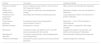
Comparison of our cases and literature findings on lupus psychosis in SLE.
| Feature | Our cases | Literature findings |
|---|---|---|
| Psychotic symptom onset | Early in disease course (Cases 1 and 2); prior to SLE diagnosis (Case 4) | Often early but can precede SLE diagnosis |
| Response to antipsychotics | Poor response, requiring immunosuppressive therapy | Resistance linked to immune and dopamine alterations |
| Presence of anti-ribosomal P antibodies | Associated with treatment-resistant psychosis | Lupus psychosis linked to anti-ribosomal P protein antibodies |
| Drug-induced psychosis | Exacerbation post-immunosuppressant discontinuation (Case 3) | Reported in ∼4.8% of SLE patients on corticosteroids |
| MRI/EEG findings | Nonspecific or normal | Often subtle or nonspecific white matter changes |
| Biomarkers | Positivity for ANA, anti-dsDNA, NLR, and CRP levels associated with SLE activity. | Important biomarkers include ANA, anti-dsDNA, anti-ribosomal P antibodies. |
| Management approach | Combined corticosteroids, immunosuppressants, and antipsychotics | Recommended multidisciplinary approach |
| Treatment challenges | Distinguishing lupus psychosis from drug-induced psychosis | Ongoing diagnostic challenge |
ANA: antinuclear antibody; BPD: bronchopulmonary dysplasia; dsDNA: double-stranded deoxyribonucleic acid; NLR: neutrophil-to-lymphocyte ratio; MRI: magnetic resonance imaging, SLE: systemic lupus erythematosus.
Anti-ribosomal P protein antibodies are more prevalent in pediatric-onset SLE compared to adult-onset SLE, with studies showing 26.7% in pediatric patients versus 6.5% in adults9 and 42% versus 7.7%, respectively.9,10 Additionally, younger adults (mean age 33.9 years) are more likely to test positive than older adults (mean age 45.3 years).10 These findings suggest that pediatric-onset SLE patients are more likely to test positive for anti-RibP antibodies, although differences in assay methods and RibP antigens used may contribute to these variations. The sensitivity and specificity of anti-RibP testing may differ between age groups, and further studies are needed to assess the diagnostic accuracy of these antibodies in both populations.
Moreover, the potential participation of antiphospholipid antibodies (aPL), these autoantibodies have been well-documented as key contributors to the pathogenesis of focal neurological manifestations in NPSLE, including headache, stroke, and epilepsy. They activate endothelial cells, platelets, and monocytes, leading to prothrombotic effects, which can result in cerebrovascular events. Additionally, aPL antibodies have been linked to diffuse neurological syndromes, such as cognitive dysfunction and seizures, suggesting that they may have pathogenic effects beyond their known role in thrombosis.11 The association of aPL antibodies with cerebrovascular disease and cognitive impairment in NPSLE emphasizes the need for further investigation into their role in disease progression and the potential for aPL as biomarkers for guiding treatment and prognosis.11
The third case demonstrated psychotic exacerbation after discontinuation of immunosuppressive therapy, aligning with studies reporting psychosis in 4.8% of SLE patients receiving corticosteroids, which often resolves upon dose reduction.1 However, in our case, psychotic symptoms persisted, necessitating combined immunosuppressive and psychiatric treatment. The fourth case, where psychotic symptoms preceded SLE diagnosis by four years, underscores the diagnostic challenge in distinguishing primary psychotic disorders from NPSLE, a phenomenon well-documented in literature.12 As neuropsychiatric symptoms in SLE often overlap with psychiatric conditions, further investigation is needed to elucidate the molecular pathways involved and to develop more targeted biomarkers for early diagnosis and management.
Imaging findings in our cases were largely nonspecific, consistent with previous studies indicating that MRI and EEG findings in NPSLE are often subtle or normal.1,12 Biomarker analysis, including ANA, anti-dsDNA, and anti-ribosomal P antibodies, remains a crucial yet nonspecific tool for diagnosis and treatment monitoring.1,12 Notably, our cases reinforce the necessity of a multidisciplinary approach, particularly in distinguishing lupus psychosis from drug-induced psychosis, an ongoing challenge in clinical practice.
ConclusionLupus psychosis is a complex manifestation of SLE that requires multifaceted treatment strategies. Our study highlights the importance of recognizing the role of immune responses and specific antibodies in treatment resistance. Future research should focus on developing targeted therapies that address both the immunological and neuropsychiatric aspects of lupus psychosis. Close interdisciplinary collaboration is crucial for effective management and improving patient outcomes.
Informed consentInformed consent was obtained from the participants to participate in the current study.
Ethical approvalThis study was conducted University of Health Sciences Erenkoy Mental Health and Neurological Diseases Training and Research Hospital and the ethical approval was waived for case reports and case series by the polices of the hospital.
FundingThis research received no specific grants from any funding agency in the public, commercial, or not-profit-sectors.
Conflict of interestThe authors declare no conflicts of interest in preparing this paper.
Data availabilityThe datasets generated during and/or analyzed during the current study are available from the corresponding author on reasonable request.


