Journal Information
Visits
6
Vol. 21. Issue 3.
(March 2025)
Images in Clinical Rheumatology
Full text access
Giant cell tumor of the tendon sheaths
Tumor de células gigantes de las vainas tendinosas
Visits
6
Carmen Raya-Santosa,
, José Antonio Bernala, José Rosasa, María Cabezas Macíanb
a Sección de Reumatología, Hospital Marina Baixa, Villajoyosa, Alicante, Spain
b Servicio de Anatomía Patológica, Hospital Universitario San Juan de Alicante, Alicante, Spain
This item has received
Article information
Full Text
Bibliography
Download PDF
Statistics
Figures (4)
Show moreShow less
Full Text
Presentation of the clinical case

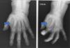
A 78-year-old woman presented with a tumour of 3 months of evolution, with progressive increase in size, on the anterior aspect of the left hand at the level of the index finger, Physical examination showed a soft, painless tumour of over 1cm in size.

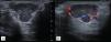
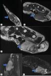
Plain radiographs (Fig. 1), ultrasound (Fig. 2), and magnetic resonance imaging (Fig. 3) were requested.

Fig. 1.

Plain radiographs in AP projections. Increased density in soft tissues (arrow) is observed.

Fig. 2.

Ultrasound of the hypoechoic, multilobed, well-defined, heterogeneous lesion (A) and with Doppler flow (B).

Fig. 3.

Resonance. Presence of hypointense lesion in sagittal and axial T1 (A, B) sections, and hyperintense in T2 axial section (C) and sagittal and axial STIR sections (D, E).

Diagnosis and evolution

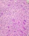
A complete excision of the lesion was performed, after which the pathological anatomy revealed a giant cell tendon sheath tumour (GCTST) (Fig. 4). Currently, the patient has not had any recurrence, with mobility preserved and no complications after the intervention.

Fig. 4.

Pathological anatomy of giant cell tumor of the tendon sheath. The black arrow points to an epithelioid macrophage and the white arrow points to an example of a giant multinucleated cell. HE, 40×.

Comment/discussion

GCTST is the second most common benign neoplasm at this level. It is classified into 2 types, depending on the involvement: localised, more frequent in hands; and diffuse. The mean age of presentation is 35–50 years, with a predominance in women. The most frequently affected location is the 2nd and 3rd fingers, and the extensor tendons, and it is also more frequent in the dominant hand.1–4

Clinically, it manifests as a solitary soft tissue nodule, asymptomatic, and with slow growth. The differential diagnosis is reached mainly with entities such as: ganglion, cyst, lipoma and sarcoma.5,6

The ultrasound description of the GCTST is a hypoechoic, heterogeneous lesion, with Doppler, related to the affected tendon. On MRI it presents as a lobed lesion, of intensity in T1 and T2 which, in STIR, show intense enhancement. Determination of hypointense haemosiderin foci is useful in differentiating these from other lesions.

With respect to histological characteristics, the GCTST presents mononuclear cells such as fibroblasts, epithelioid or foamy macrophages; multinucleated giant cells and inflammatory cells, on collagen stroma. On the other hand, vascularisation can be observed, with hemosiderin deposition in some cases.3,7,8

Treatment is based on the total removal of the lesion, although up to 45% recur.3

Ethical responsibilities

Protection of people and animals. The authors state that no experiments were carried out on humans or animals for this research.

Data confidentiality. The authors state that they have followed their workplace's protocols as regards the publication of patient data.

Right to privacy and informed consent. The authors obtained informed consent from the patients or subjects referred to in the article. This document is in the possession of the corresponding author.

Declaration of competing interest

None.

References
[1]
A. Orois Codesal, J.A. Oteo Maldonado, P. Benavente.
Tumor de células gigantes de la vaina tendinosa de la mano. Estudio de la epidemiología, eficacia de las pruebas de imagen en su diagnóstico y análisis de la recidiva.
Rev Iberoam Cir Mano., 44 (2016), pp. 8-12
[2]
M. Lautenbach, S. Kim, M. Millrose, A. Eisenschenk.
Nodular giant cell tumour of the tendon sheath of the hand: analysis of eighty-four cases: diagnostic decisions and outcome.
Int Orthop., 37 (2013), pp. 2211-2215
[3]
S. Stacchiotti, H.R. Dürr, I.M. Schaefer, K. Woertler, R. Haas, A. Trama, et al.
Best clinical management of tenosynovial giant cell tumour (TGCT): a consensus paper from the community of experts.
Cancer Treat Rev., 112 (2023),
[4]
E. Villanueva García, M. Díez Risueño.
Tumor de células gigantes localizado en la falange proximal del pulgar. Caso clínico y revisión bibliográfica.
Rev Esp Cir Ortopédica Traumatol., 47 (2003), pp. 138-140
[5]
J. Ho, T. Peters, B.C. Dickson, D. Swanson, A. Fernandez, A. Frova-Seguin, et al.
Detection of CSF1 rearrangements deleting the 3’ UTR in tenosynovial giant cell tumors.
Genes Chromosomes Cancer., 59 (2020), pp. 96-105
[6]
M. Md, R. Jh, L. Rb, F.S. Jc, F. Dj, W. Ea.
Pigmented villonodular synovitis: radiologic-pathologic correlation.
Radiogr Rev Publ Radiol Soc N Am Inc., 28 (2008), pp. 1493-1518
[7]
M. Díaz Sánchez, A. Vadillo Santos, M.J. Becerril Barrientos, Mde G. Gómez Pérez.
Tumor de células gigantes de la vaina tendinosa.
Acta Médica Grupo Ángeles., 20 (2022), pp. 197-198
[8]
B. López Ruiz, J. Gasch Blasi, A. Rodríguez Fernández.
Tumor de células gigantes de la vaina tendinosa en el seno del tarso.
Rev Pie Tobillo., 25 (2011), pp. 27-30
Copyright © 2025. Sociedad Española de Reumatología (SER), Colegio Mexicano de Reumatología (CMR) and Elsevier España, S.L.U.
Download PDF
Idiomas
Reumatología Clínica (English Edition)
Article options
Tools
es en

¿Es usted profesional sanitario apto para prescribir o dispensar medicamentos?

Are you a health professional able to prescribe or dispense drugs?