A 65-year-old woman came to the emergency department for continuous pain and swelling at the ulnar dorsal margin of the right fifth metacarpophalangeal joint which had lasted 4 days. The patient had no other signs in the rest of the joints of the hands or the rest of the skeleton.
At the onset of symptoms, the patient went to her family physician, who prescribed antibiotics, but after noting no improvement, decided to visit the emergency room. Upon interrogation she reported no known diseases or history of trauma, nor had fever. In the absence of history and because of the monoarticular involvement, the emergency room doctor decided to rule out an infectious origin, requesting an ultrasound guided arthrocenthesis for microbiology analysis.
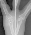
The ultrasound showed no effusion, but found a soft tissue injury, partially defined, and heterogeneous echotexture with hyperechoic areas. The tumor was in contact with the cortical bone of the metacarpal, which remained intact, except for an area where we observed a marked erosion, with a soft tissue lesion projecting into the medullary cavity (Fig. 1). Given the ultrasonography findings, X-rays were analyzed, confirming increased periarticular soft tissues, with very high density, accompanied by erosion of the metacarpal head, which as a hallmark had a sclerotic margin (Fig. 2). These sonographic and radiographic findings indicated periarticular tophi associated with bone erosion. The ultrasound-guided arthrocenthesis of the tophus showed a dense whitish material that under polarized light microscopy was identified as monosodium urate crystals (Fig. 3).
X-rays centered on the fifth metacarpophalangeal joint of both hands that allows for the assessment of the bone erosion comparatively and shows the sclerotic margin (arrows) with soft tissue augmentation and high density in the medial aspect of the right metacarpal head. There was no involvement of the joint space.
During the initial clinical examination, gout was not considered because the involvement of the hand is not usual (more often it occurs feet-first on the metatarsophalangeal-tarsal area followed by ankles and knees) and because the patient did not present any of the conditions (alcoholism, diabetes mellitus, hypertension, renal disease or diuretic use) frequently associated with gout. In addition, in Emergency departments, infectious arthritis is considered as the prototype of acute monoarticular disease versus other common diseases such as gout, which typically is characterized by recurrent attacks of monoarthritis.1–3
Ethical ResponsibilitiesProtection of People and AnimalsThe authors state that no experiments were performed on humans or animals.
Data ConfidentialityThe authors state that no patient data appear in this article.
Right to Privacy and Informed ConsentThe authors state that no patient data appear in this article.
Conflict of InterestThe authors have no disclosures to make.
Please cite this article as: Moran LM, Martinez LP. Ataque agudo de gota sospechoso de artritis infecciosa. Reumatol Clin. 2013;9:324–325.


