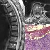
A 76-year-old female patient came to the clinic due to lumbar spine pain and weight loss, which had progressed over the past 6 months. She had been diagnosed with systemic sclerosis (SS) and interstitial lung disease 5 years prior and had been treated with cyclophosphamide induction therapy for 18 months (6 monthly cycles and 4 trimestral ones), followed by azathioprine and low dose steroid as maintenance treatment. Upon examination, the patient presented cachexia and diffuse pain on spinous process palpation. Spinal X-rays showed anterior vertebral body collapse at D8 and a magnetic resonance (MR) revealed multiple round lesions with a fluid-fluid level, with a hyperintense signal on the superior level potentiated sequence on T2 and an intermediate signal on T1, with hypointense signal on the inferior level of both sequences (Fig. 1A and B). A bone biopsy revealed that the lesions were metastasis from a poorly differentiated signet ring cell adenocarcinoma, surrounding hemorrhagic content (Fig. 1C). From an immunohistochemical point of view, the tumor cells expressed cytokeratin 7 and protein 15 of cystic disease liquid, indicating a likely breast cancer origin. However, no primary tumor was found in follow-up, which included a bilateral mastorgraphy, a craneo-cervical and thoraco-abdomino-pelvic computed tomography, a complete radionucleide bone scan, an endoscopy and a colonoscopy. The patient was treated with paclitaxel plus carboplatin, third level analgesia, zolendronate and palliative radiotherapy, remaining stable for 6 months afterward.
Dorsolumbar spine magnetic resonance showing, in sagital (A) and axial (B) projections of a T2 potentiated sequence, the presence of multiple vertebral lesions with typical fluid–fluid levels, hyperintense superiorly and hypointense inferiorly. A bone biopsy (C) revealed that these lesions corresponded to metastasis of a poorly differentiated adenocarcinoma with signet ring cells (arrowheads), surrounding a hemorrhagic content (arrows) (hematoxilin-eosin 10×).
Fluid–fluid levels may appear when substances with different densities are contained within a cystic or compartmentalized structure.1 The presence of these levels in a bone lesion suggests internal bleeding, followed by stratification of the blood components in relation to their density.1–3 These levels may be seen on X-rays or tomography, but the best detection method is MR.3 The superior level is usually hyperintense in relation to the inferior one in the T2 potentiated sequence because the latter may contain cellular components. However, in the T1 potentiated sequence the aspect varies depending on the stage of the lesion, showing an increased signal in the superior level when the hemorrhage is subacute, presumably due to the presence of extracellular metahemoglobin.4 Fluid–fluid levels have been detected in various benign bone lesions, oncluding aneurysmatic bone cysts, simple bone cysts, giant cell tumors, chondroblastoma, osteoblastoma, brown tumors, fibrous dysplasia, Langerhans’ hystiocytosis, intraosseous ganglions and hemangiomas, but also in malignant lesions such as telangiectasic osteosarcoma, the malignant fibrous hystiocytoma, fibrosarcoma, and plasmocytoma.1,3 However, these levels appear rarely in bone metastasis, with only 6 cases published up to date.2,3,5–8 In 3 of them there was a past history of cancer, breast2,5 and bladder.7 However, in the rest of the cases, the detection of fluid-fluid levels revealed the presence of neoplasia, 2 of them respiratory in origin6,8 and one unknown,3 as our patient's case. The lesions were multiple in 3 cases, all of them affecting the spine.2,3,7 Although the history of SS probably had no influence on the type of metastasis, these patients have an increased risk of neoplasia, especially lung, hematological, bladder and hepatocellular,9 with diagnosis not always easy in early stages. Another factor that must be taken into account in this case is treatment with cyclophosphamide because this drug may be associated with neoplasias such as leukemia, lymphoma, non-melanoma skin cancer and bladder neoplasia.10 In conclusion, although fluid-fluid levels are not specific, when faced with the presence of multiple vertebral lesions of this kind, it is important to consider bone metastasis among the differential diagnosis.
Ethical ResponsibilitiesProtection of persons and animalsThe authors state that no experiments were performed on persons or animals for this study.
Data confidentialityThe authors state that they have followed their workplace protocols regarding the publication of patient data and all patients included in the study have received enough information and have given their written informed consent to participate in the study.
Right to privacy and informed consentThe authors state that they have obtained informed consent from patients and/or subjects referred to in this article. This document is in the possession of the corresponding author.
Conflict of InterestThe authors deny any conflict of interest.
Please cite this article as: Sifuentes Giraldo WA, Pachón Olmos V, Gallego Rivera JI, Vázquez Díaz M. Metástasis vertebrales múltiples con niveles líquido-líquido. Reumatol Clin. 2013;10:338–339.


