Journal Information
Vol. 9. Issue 4.
Pages 250-251 (July - August 2013)
Visits
7202
Vol. 9. Issue 4.
Pages 250-251 (July - August 2013)
Images in Clinical Rheumatology
Full text access
Osteomalacia Produced by a Hyperphosphaturic Tumor
Osteomalacia provocada por un tumor hiperfosfatúrico
Visits
7202
Miguel Cantalejo Moreiraa,
Corresponding author
, Raúl M. Veiga Cabelloa, José M. Ruiz Giardínb
a Unidad de Reumatología, Hospital Universitario de Fuenlabrada, Madrid, Spain
b Servicio de Medicina Interna, Hospital Universitario de Fuenlabrada, Madrid, Spain
This item has received
Article information
Full Text
Bibliography
Download PDF
Statistics
Figures (3)
Show moreShow less
Full Text

Oncogenic osteomalacia is caused by the action of fibroblast growth factor 23 (FGF 23) on the renal proximal tubule, causing hyperphosphaturia and, as a consequence, bone hypophosphatemia and metabolic disease.1,2 FGF 23 usually occurs in tumors of mesothelial origin.3 Less than 150 cases are described in the literature.

Case

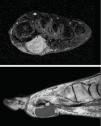
A 53 year woman, without notable history, came to the emergency room presenting mechanical, intense pelvic pain and inability to stand or walk. Urgent laboratory analysis highlighted an alkaline phosphatase value of 398U/l (30–120), the rest of the blood count and biochemistry values were normal. AN AP radiograph of the pelvis showed osteopenia with no other findings. The patient was admitted and a second set of laboratory tests requested: phosphate 1.2mg/dl (2.5–4.5), alkaline phosphatase 395U/l (30–120), 1.25 vitamin D3 32.7ng/ml (30–40), phosphaturia, 1436.00mg/24h (400–1300). Other values were normal. An isotope bone scan was requested (Fig. 1), as were a pelvic CT (Fig. 2) and a pelvic MRI. A subsequent physical examination demonstrated that the patient had hard, painless, 2cm×2cm erythematous and bullous lesions on the sole of her left foot. Suspecting a phosphaturic tumor an octreotide scintigraphy was requested and found normal. We requested an MRI of the foot (Fig. 3) and removed the lesion. The pathologic diagnosis was: mixed connective tissue variant mesenchymal tumor. After removal of the tumor, the patient returned to normal blood and urine phosphate values and no new fractures occur.

Fig. 1.

Bone scan: multiple bone involvement, with fixed tracer (99mTc-diphosphonates) found in bilateral ribs, a vertebral body, the horizontal branch of the pubis and right inferior pubic ramus.

(0.22MB).
Fig. 2.

CT scan of pelvis showing multiple fractures in different stages of progression at the level of the right pubis and ischium bilaterally.

(0.15MB).
Fig. 3.

MRI of left foot: soft tissue solid tumor, poly-lobular, measuring 5.3cm×2.6cm×2cm in a subdermal location, inserted deep into the muscular plane, separated from the bone.

(0.22MB).
Discussion

The first case was described by Mc Cance in 19474 and the association of the tumor with metabolic disease was made in 1959 by Prader et al.5 Less than 150 cases are described in the literature. Most tumors are of mesenchimal origin,6,7 with many cases being small and difficult to locate tumors, a situation that delays diagnosis 2.5 years (2.5 months–19 years) on average from the onset of locomotor symptoms.8 Many of these tumors express somatostatin receptors, therefore labeled octreotide scintigraphy may be useful in the diagnosis.8,9 Removal of the tumor often resolves the metabolic8 disorder.

Ethical disclosures

Protection of human and animal subjects. The authors declare that the procedures followed were in accordance with the regulations of the responsible Clinical Research Ethics Committee and in accordance with those of the World Medical Association and the Helsinki Declaration.

Confidentiality of Data. The authors declare that no patient data appears in this article.

Right to privacy and informed consent. The authors must have obtained the informed consent of the patients and /or subjects mentioned in the article. The author for correspondence must be in possession of this document.

References
[1]
R. Kumar.
Tumor-induced osteomalacia and the regulation of phosphate homeostasis.
Bone, 27 (2000), pp. 333-338
[2]
A.E. Nelson, R.C. Bligh, M. Mirams, A. Gill, A. Au, A. Clarkson, et al.
Fibroblast growth factor 23: a new clinical market for oncogenic osteomalacia.
J Clin Endocrinol Metab, 88 (2003), pp. 4088-4094
[3]
J.L. Dupond, H. Mahammedi, D. Prie.
Oncogenic osteomalacia: diagnostic importance of fibroblast growth factor 23 and F-18 fluorodeoxyglucose PET/CT scan for the diagnosis and follow-up in one case.
[4]
R.A. Mc Cance.
Osteomalacia with Looser's nodules due to a raised resistance to vitamin D acquired about the age of 15 years.
Q J Med, 16 (1947), pp. 33-46
[5]
V. Prader, R. Illig, E. Vehlinger, G. Stalder, Rachitis.
Infolge, knochen tumors.
Helv Paediatr Acta, 14 (1959), pp. 554-565
[6]
S.M. Jan De Beur.
Tumor induced osteomalacia.
Primer on the metabolic bone diseases and disorders of mineral metabolism, 6th ed., pp. 345-350
[7]
M.K. Drezner.
Tumor-induced osteomalacia.
Rev Endocr Metab Disord, 2 (2001), pp. 175-186
[8]
A.L. Folpe, J.C. Fanburg-Smith, S.D. Billings, M. Bisceglia, F. Bertoni, J.Y. Cho, et al.
Most osteomalacia associated mesenchymal tumors are a single histopathologic entity: an analysis of 32 cases and a comprehensive review of the literature.
Am J Surg Pathol, 28 (2004), pp. 1-30
[9]
C. Garcia, R. Spencer.
Bone and In-111 octeotride imaging in oncogenic osteomalacia: a case report.
Clin Nucl Med, 27 (2002), pp. 582-583

Please cite this article as: Cantalejo Moreira M, et al. Osteomalacia provocada por un tumor hiperfosfatúrico. Reumatol Clin. 2013;9:250–1.

Copyright © 2012. Elsevier España, S.L.. All rights reserved
Download PDF
Idiomas
Reumatología Clínica (English Edition)
Article options
Tools
es en

¿Es usted profesional sanitario apto para prescribir o dispensar medicamentos?

Are you a health professional able to prescribe or dispense drugs?