


The use of well characterized osteoarthritis (OA) cohorts is mandatory for the study and knowledge of this disease. Currently, there is no prospective cohort in this pathology in Spain. The objective of this work is to describe the first osteoarthritis cohort in Spain, PROCOAC (Cohort PROspectiva de A Coruña).
MethodsThe Unit of Rheumatology of the University Hospital of A Coruña started a prospective follow-up study in 2006. The patient inclusion criteria were: I) Patients older than 55 years who underwent an abdominal x-ray to study both hips II) Patients diagnosed with radiographic hand OA according to ACR criteria III) Patients diagnosed with radiographic knee or hip OA according to ACR criteria. Follow-up was performed every two years collecting clinical, analytical, genetic and radiographic information.
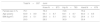
ResultsThe cohort consists of 937 patients, 873 have radiographic knee OA, 783 hip OA and 679 hand OA. The mean age of the population is 63.9±8.9 years and the average BMI is 29.6±5.1. More than half of the population has high blood pressure and 17% diabetes. The predominant osteoarthritis in the hand is nodular (78.1%), followed by trapeziometacarpal (55.3%) and erosive (18.4%). Twenty-one point four percent and 43.1% are healthy at knee and hip level respectively; observing a grade 1 in 26% and 37%; a grade 2 in 26.7% and 11.5%; a grade 3 in 14.9% and 4%; and a grade 4 in 9.4% and 3.7% respectively. Of the population, 44.1% has only 1 joint affected, 39.9% has 2 and 13.4% has 3 joints affected. Age (OR=1.11; p<.001), BMI (OR=1.11; p=.002) and total WOMAC (OR=1.03; p=.005) are the only risk factors if we compare the involvement of a single location versus three. A discrepancy between pain and radiographic damage at the joint level was also detected in patients with KL≤2 grade, and therefore a significantly higher percentage of patients with knee OA experienced pain (66.1%) compared to patients with OA hip (21.1%) (p<.001).
ConclusionsThe PROCOAC cohort is an instrument that allows studies of incidence and progression in hand, knee and hip OA; as well as determining factors that are associated with the different OA phenotypes.
El uso de cohortes de Osteoartritis (OA) bien caracterizadas es obligatorio para estudiar y profundizar en el conocimiento en esta enfermedad. En España no existe actualmente ninguna cohorte prospectiva en esta patología; así el objetivo de este trabajo es describir la primera cohorte de Osteoartritis en España, la PROCOAC (PROspective COhort of A Coruña).
Material y métodosEl Servicio de Reumatología del Hospital Universitario de A Coruña inició un estudio de seguimiento prospectivo en el año 2006. Los criterios de inclusión fueron: I) Pacientes mayores de 55 años a los que se les realizó una radiografía abdominal que permitiese estudiar ambas caderas II) Pacientes diagnosticados de OA radiográfica de mano según los criterios ACR III) Pacientes diagnosticados de OA radiográfica de rodilla y/o cadera según los criterios ACR. Se realizó seguimiento cada dos años recogiendo información clínica, analítica, genética y radiográfica.
ResultadosLa cohorte consta de 937 individuos, 873 tienen OA radiográfica de rodilla, 783 de cadera y 679 de mano. La edad media de la población es 63,9±8,9 años y el IMC promedio de 29,6±5,1. Más de la mitad de la población tiene hipertensión arterial y el 17% diabetes. La osteoartritis predominate en la mano es la nodular (78,1%), seguida de la rizartrosis (55,3%) y la erosiva (18,4%). El 21,4% y el 43,1% son sanos a nivel de rodilla y cadera respectivamente; observando un grado 1 en el 26% y 37%; un grado 2 en el 26,7% y 11,5%; un grado 3 en el 14,9% y 4%; y un grado 4 en el 9,4% y 3,7% respectivamente. El 44,1% de la población tiene 1 articulación afectada, el 39,9% tiene 2 y el 13,4% tiene 3 articulaciones afectadas. La edad (OR=1,11; p<0,001), el IMC (OR=1,11; p=0,002) y el WOMAC total (OR=1,03; p=0,005) son los únicos factores de riesgo si comparamos la afectación de una sola ubicación frente a tres. También se detectó una discrepancia entre el dolor y el daño radiográfico a nivel articular en pacientes con grado KL≤2, de modo que un porcentaje significativamente mayor de pacientes con OA de rodilla experimentaron dolor (66,1%) en comparación con pacientes con OA de cadera (21,1%) (p<0,001).
ConclusiónLa cohorte PROCOAC es un instrumento que permite realizar estudios de incidencia y pro-gresión en la osteoartritis de mano, rodilla y cadera, así como conocer factores que se asocian con losdiferentes fenotipos de osteoartritis.
Osteoarthritis (OA) is the most common musculoskeletal disease.1 Until very recently it was defined as a purely degenerative process which led to the degradation and destruction of joint cartilage. However, in recent years, thanks to studies conducted on the disease, it has been confirmed that the process which takes place and which triggers the disease is highly active, due to the imbalance between joint tissue destruction and repair.2
OA is also considered to be a disease of the whole diarthrodial joint which is currently defined as an organ (the combination of specialized tissues which possess a certain structure and organisation for meeting with a specific function).3 The trigger for the disease is usually damage brought about by one of the components involved (joint cartilage, synovial membrane, bone tissues, intra and periarticular soft tissues), which leads to joint failure. Furthermore, it is characterised by the progressive degradation and loss of the hyaline cartilage and subchondral bone, in addition to damage which occurs in the synovial tissues, associated with a thickening and sclerosis of the subchondral film, the formation of osteophytes, the distention of the joint capsule and changes to the periarticular soft tissues.4
All of these conceptual changes are possible, due to the continuous study in the field of medicine and more specifically in that of rheumatic diseases. The need to continue conducting studies in the population is imperative in order to determine new approaches to the concept, classification, diagnosis and treatment of this entity. To do so, the creation and follow-up of patient cohorts may be of significant contribution.
Against this backdrop, for further study of this disease, in the year 2006 we initiated the creation and follow-up of a cohort of patients diagnosed with OA, called PROCOAC (PROspective COhort of A Coruña) in our centre. This enabled us to carry out incidence and progression studies that are associated with different OA phenotypes. In this article we describe the methodology used for its constitution, together with demographic characteristics and main clinical traits.
Material and methodsPatients (PROCOAC cohort)The PROCOAC cohort focuses on the study of peripheral arthritis of the hands, knees and hips. This project started with the study of a group of patients who attended the Emergency Services of our hospital due to abdominal pain and who had an X-ray of the abdomen where we were able to assess both hips in our population. With that information we also studied the presence of hip osteoarthritis. Thus, the inclusion criteria of the population making up the cohort were: a) patients over 55 who attended the Emergency Services of our hospital and those who had undergone a plain X-ray of the abdomen (including both hips, which allowed us to detect the presence or absence of osteoarthritis); b) patients who attended our Rheumatology Service with pain in their hands and with a radiographic diagnosis of OA in keeping with the American College of Rheumatology (ACR)5 criteria; c) patients with knee pain and with a radiographic OA diagnosis of the knee in keeping with the ACR6 criteria and patients with hip pain and diagnosed radiographically with OA of the hip, also in keeping with ACR7 criteria.
This inclusion process began in the year 2006 and remains active to date. All patients signed an informed consent form approved by the local ethics committee. After this, data were collected (review of medical record and interview with the patient) to complete the medical history (demographic and clinical information), biological samples (general analysis and genetic studies) and X-rays of both hands, knees and hips were taken.
The recorded variables included a complete medical history (personal and family background), review of comorbidities (psoriasis, high blood pressure, diabetes, dyslipidaemia), a physical examination (anthropometry and joint examination). To measure outcomes, we used assessment scales for the 3 joint groups (visual analogous scales for physician and patient, AUSCAN, FIHOA, HAQ, WOMAC, Lequesne of the knee and hip, HAD and SF-12).2,8–15
All procedures were made at the first visit and repeated in those established for follow-up every 2 years.
The radiologic grade was assessed for all participants, following the Kellgren-Lawrence16 scale for classifying OA of the knee and hip and also the presence of joint prosthetics or fracture (including date and cause).
To assess the hand, information referring to the type of condition was collected: trapeziometacarpal osteoarthritis (rhizarthrosis), nodular osteoarthritis (number of nodules and location), erosive osteoarthritis (number of erosions and location), osteoarthritis in metacarpophalangial joints or inflammation. These different phenotypes may coexist in the same patient.
To assess the severity of the disease, the number of joints affected was coded as 0–1 and 2–3, in keeping with the radiographic information of the hands, knees and hips (Table 1).
All data was introduced into a notebook for electronic data collection called SIMON (for its initials in Spanish), an intelligent monitoring system which, specifically for this OA cohort, consisted of 869 variables (151 as the base register and 718 for each follow-up).
Statistical analysisStatistical analysis was performed using the SPSS v. 24, and included contingency χ2 tables and binary multivariate logistic regression models, analysing the influence of different variables as potential risk and confusion factors, such as age, sex and body mass index (BMI). Using this model we explored the possible associated risk factors for polyarticular involvement of this entity.
ResultsCohort descriptionAt the time of analysis, in the year 2018, the cohort consisted of 937 people, 873 (93.1%) with radiographic OA of the knee, 783 (83.5%) with radiographic OA of the hip and 679 (72.4%) with radiographic OA of the hand. The mean age of the population was 63.9 years (±8.9 years) and average BMI 29.6 (±5.1). The great majority were women (75.1%) and 29% were smokers (active or ex-smokers) (Table 2). The percentage of follow-up losses was 19.01%.
With regard to the clinical variables, over half of the population had high blood pressure (55.2%) and hypercholesterolaemia (54.2%), 16.3% diabetes mellitus and 18.5% psoriasis (personal involvement or family history in first grade) (Table 3).
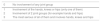
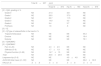
Analysis of the analytical variables showed that 11.2% and 2.2% had a positive rheumatoid factor and ACPA, respectively, and the acute phase reactant levels were within the parameters of normality (Table 4).
Analytical variables of PROCOAC.
| Total N=937 | Joint | |||
|---|---|---|---|---|
| Knee N=873 | Hip N=783 | Hand N=679 | ||
| ESR (mmHg) | 20.5±14.7 | 20.8±14.8 | 20.9±14.9 | 20.7±15.1 |
| PCR (mg/dL) | .5±1.6 | .5±1.6 | .5±1.7 | .5±1.7 |
| RF (positive) | 11.2% | 11.2% | 11.8% | 11.1% |
| Ac CCP (positive) | 2.2% | 2.2% | 2.3% | 2.2% |
Ac CCP: antibodies against cyclic citrullinated peptides; ESR: erythrocyte sedimentation rate; RCP: reactive C protein; RF: rheumatoid factor.
Analysis of radiographies show that 43.1% of the population was Grade 0 in the hip and 21.4% in the knee (Table 5). 26% had Grade 1 in the knee and 37% in the hip; 26.7% had a Grade 2 in the knee and 11.5% in the hip; 14.9% had a Grade 3 in the knee and 4% in the hip. However, 9.4% and 3.5% were Grade 4 on the Kellgren and Lawrence (KL) scale in the knee and hip, respectively. 1.6% of patients had a knee replacement and .6% had a hip replacement.
PROCOAC joint assessment.
| Total N=937 | Joint | |||
|---|---|---|---|---|
| Knee N=873 | Hip N=783 | Hand N=679 | ||
| [0,1–5]KL grading in % | ||||
| Grade 0 | NE | 21.4 | 43.1 | NE |
| Grade 1 | NE | 26.0 | 37.0 | NE |
| Grade 2 | NE | 26.7 | 11.5 | NE |
| Grade 3 | NE | 14.9 | 4.0 | NE |
| Grade 4 | NE | 9.4 | 3.7 | NE |
| Prothesis | NE | 1.6 | .6 | NE |
| [0,1–5] | ||||
| [0,1–5]Type of osteoarthritis in the hand in % | ||||
| Trapeziometacarpal | NE | NE | NE | 55.3 |
| Erosive | NE | NE | NE | 18.4 |
| Nodular | NE | NE | NE | 78.1 |
| [0,1–5]WOMAC | ||||
| [0,1–5]WOMAC | ||||
| Pain (0−20) | NE | 4.3±4.5 | NE | NE |
| Stiffness (0−8) | NE | 1.7±1.9 | NE | NE |
| Physical function (0−68) | NE | 14.7±15 | NE | NE |
| Total | NE | 20.6±20.5 | NE | NE |
| Lequesne hip (0−8) | NE | NE | 4.9±4.8 | NE |
| AUSCAN total mean (0−100) | NE | NE | NE | 36.2±30.5 |
| HAQ (0−3) | .5±.6 | .5±.6 | .5±.6 | .5±.6 |
AUSCAN: Australian-Canadian Osteoarthritis Hand Index; HAQ: Health assessment questionnaire; KL: Kellgren and Lawrence; NE: non-evaluable; WOMAC: Western Ontario and McMaster Universities Arthritis Index.
The most frequent OA of the hands was the nodular phenotype (78.1%) rhizarthrosis was present in 55.3% and the erosive OA of the hands in 18.4%.
A discrepancy was detected between pain and radiographic damage in knees or hips in patients with Grade KL≤2, so that a significantly higher percentage of patients with OA of the knee experienced pain (66.1%) compared with patients with OA of the hip (21.1%) (P <.001).
Polyarticular osteoarthritisThe percentage of patients with radiographic OA in more than one joint was 53.3% (2 joints 39.9% and 3 joints 13.4%), whilst involvement of 0–1 joints was present in 46.7% (one in 44.1%).
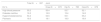
When they were compared in a binary logistic regression analysis to confirm whether there was a causal relationship in patients with a single affected joint to patients with 3 locations we found that the factors associated with polyarticular OA were age (OR=.,11; 95% CI: 1.06–1.16; p<.001), BMI (OR=1.11; 95% CI:1.04−1.19; p=.002) and total WOMAC (OR=1.03; 95% CI:1.01–1.05; p=.005), without a specific inflammatory component in patients with 3 locations (Table 6). For this, we took into account the different confusion factors, such as age, sex and BMI.
Logistic regression model to analyse the variables associated with the number of affected locations (one location compared with 3 locations).
| OR | 95% CI | P | ||
|---|---|---|---|---|
| Low | High | |||
| Female | .96 | .38 | 2.45 | .31 |
| Age | 1.11 | 1.06 | 1.16 | <.001* |
| BMI | 1.11 | 1.04 | 1.19 | .002* |
| WOMAC total | 1.03 | 1.01 | 1.05 | .005* |
| PCR | .98 | .81 | 1.18 | .836 |
| VSG | .99 | .96 | 1.01 | .274 |
| Psoriasis | .83 | .38 | 1.81 | .642 |
BMI: body mass index; CI: confidence interval; CRP: C reactive protein; ESR: erythrocyte sedimentation rate; OR: odd ratio; WOMAC: Western Ontario and McMaster Universities Arthritis Index.
Advances in OA have been appearing in recent years thanks to research carried out in the last few decades. Despite this being the most common musculoskeletal disease,1 medical and therapeutic approach has remained obsolete, since it was always classified as a degenerative disease and one of ageing: the different patterns of involvement or phenotypes were therefore relegated to a single catch-all box entitled “arthrosis”.
Very recently the concept of OA was redefined due to the fact that a highly active process in joint repair and destruction was demonstrated.2 The International Society for Osteoarthritis Research (OARSI) clearly defines that there is a background causal disorder of major cellular stress, with its consequent degradation of the extracellular matrix, where these lesions are also the response to changes of the pro-inflammatory pathways of innate immunity. In a primary stage molecular changes are observed (abnormal metabolism of joint tissues), followed by physiological and anatomical changes (degradation of cartilage, bone remodelling, formation of osteophytes, joint inflammation, loss of correct joint function) and, lastly, the appearance of pain.17
The use of well-structured cohorts followed up in time seem to be a good ally for approaching research into this disease, where the study of the disease pathogenesis and the different phenotypes of involvement may throw some light on possible therapeutic applications.
As with other events, this disease mostly affects women, 75.1% in our cohort. In the Spanish population18 it is one of the factors associated with OA of the hands and knees, with an association in the limit of statistical significance in OA of the hip.
When we speak of cohorts we must mention one of the most important records worldwide for the study of cardiovascular risk, the Framingham study initiated in 1948. However, there are now major cohorts which focus on OA, including the Cohort Hip and Cohort Knee, (CHECK, Holland) and that of the Osteoarthritis Initiative (OAI, U.S.A.).19
Regarding BMI, our population is at levels of extra weight (29.6) and obesity type 1, with no differences observed between the 3 joint groups and no major differences with the other 2 cohorts (26 in that of CHECK and 28 in that of OAI).19 This was also detected in the latest EPISER18 study as one of the factors associated with this disease in the 3 locations. The explanation of this fact is related to changes in life habits of the population in recent years, where the numbers of obese and overweight people have risen worldwide, along with tobacco consumption,18,20 the latter being present in 29% of our population.
Unlike our cohort, the origin of the subjects for both correspond in 75% to adverts, including on web sites.21 Also, the inclusion criteria are different too: that of the CHECK focuses on the knee or hip and that of the OAI on the knee, exclusively.19 In the analysis of hand involvement we included the knee and hip as well. Evaluations were performed every 2 years, although the other cohorts carried out these procedures annually.
Radiologic damage in the knees is more notable in the OAI and PROCOAC compared with that of the CHECK. Regarding the hips, we were only able to compare with the CHECK and in this case the PROCOAC also shows higher radiologic involvement.
These differences in joint involvement are not, however, reflected in the WOMAC pain, stiffness and function: they are higher in the PROCOAC and CHECK compared with the OAI.
Unlike the other 2 cohorts, in our case there were no rigorous age limits nor was any criterion established based on the radiologic grade for participation. We were more interested in the study of the diagnosis and follow-up. By contrast and defect, in our population no imaging analysis through magnetic resonance was carried out. Due to all of this we have 3 cohorts which address the same disease, but in different joint groups or with different evaluations, which present comparative limits in all areas assessed, given that they were created with different objectives.
Our cohort has several limitations. One is that it only represents patients in a hospital environment and comparison with healthy patients (very low number) is limited. Furthermore, there were few patients who sought medical attention for OA of the hip as a reason for primary care, since they usually consulted this event in another specialty, such as the trauma unit. We cannot therefore state with these data that our cohort is representative of OA in Spain, because cohort creation and design was mainly directed at the study and advance of this disease regarding the concept of the different phenotypes and therapeutic attitudes based on them.
Conflict of interestsThe authors have no conflict of interests to declare.
Please cite this article as: Oreiro-Villar N, Raga AC, Rego-Pérez I, Pértega S, Silva-Diaz M, Freire M, et al. Descripción de la cohorte PROCOAC (PROspective COhort of A Coruña): Cohorte prospectiva española para el estudio de la osteoartritis. Reumatol Clin. 2022;18:100–104.


