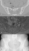
We report the case of a 50-year-old woman who presented with a 3-month history of inflammatory low back pain and arthritis of the knees, with no other type of manifestations. Laboratory analyses demonstrated an elevated C-reactive protein level (2.5mg/dL) and erythrocyte sedimentation rate (43mm/h) and she was found to be negative for human leukocyte antigen (HLA) B27; there were no other abnormal findings. In the initial radiological study, plain radiography demonstrated meniscal calcification in both knees and sclerosis in both sacroiliac joints; magnetic resonance imaging (MRI) showed bone marrow edema in a short-tau inversion recovery (STIR) sequence (Fig. 1A and B). We observed no syndesmophytes affecting the axial skeleton. These findings led us to consider a differential diagnosis including spondyloarthritis (SpA) and pyrophosphate arthropathy. There were rectangular extracellular crystals with positive birefringence in synovial fluid obtained from knee. We requested computed tomography (CT) of the sacroiliac joints (Fig. 1C), which showed sclerosis with linear intra-articular calcifications in both joints. The patient was diagnosed with arthritis caused by calcium pyrophosphate deposition. Treatment was begun with low-dose steroids and colchicine, and the symptoms improved.
(A) Anteroposterior radiograph of pelvis showing calcification in fibrocartilage in the pubic symphysis, as well as sclerosis in both sacroiliac joints with cortical irregularity. (B) Axial slice of magnetic resonance of the pelvis. Short-tau inversion recovery (STIR) sequence, with hyperintense signal in both sacroiliac joints. (C) Computed tomography, axial image, of pelvis, showing the vacuum phenomenon in right sacroiliac joint, as well as calcification in fibrocartilage of both sacroiliac joints, with osteophytes.
Chondrocalcinosis develops due to calcium pyrophosphate dehydrate crystals in the fibrocartilage of the joints.1 The joint most widely affected is the knee, although others are usually also involved. The diagnosis is based on the visualization of crystals in the synovial fluid.1
Plain radiography enables the observation of calcification in the fibrocartilage and hyaline cartilage.1
In the case of diagnostic doubt, CT can be a useful aid. The images produced by MRI are little specific.2–4 There are reports of cases of arthritis due to pyrophosphate involving the axial skeleton in the literature.5
The sacroiliac joint is not characteristically affected by diseases of this type. The type of pain—inflammatory and chronic in this case, together with the radiological changes, made it necessary to include SpA in the differential diagnosis. Edema in the sacroiliac joints is not always secondary to SpA.
Ethical DisclosuresProtection of human and animal subjectsThe authors declare that no experiments were performed on humans or animals for this study.
Confidentiality of dataThe authors declare that they have followed the protocols of their work center on the publication of patient data.
Right to privacy and informed consentThe authors declare that no patient data appear in this article.
Conflict of InterestThe authors declare they have no conflicts of interest.
Please cite this article as: Moreno Martinez MJ, Moreno Ramos MJ, Linares Ferrando LF. Sacroilitis por pirofosfato cálcico. Reumatol Clin. 2018;14:175–176.


