Hyperlipidaemia can present accompanied by tendon xanthomatosis. Xanthomas are clusters of collagen and cholesterol-filled macrophages. The Achilles tendon, extensor tendons of the hand, and the elbow tendons are principally affected. Joint ultrasound can be a very useful tool for diagnosing and monitoring the course of tendon xanthomas. Clinicians usually request screening for xanthoma in patients with suspected family hypercholesteraemia (FH), since the presence of xanthomas forms part of the diagnostic criteria of the disease.
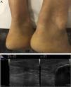
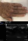
Presentation of the Clinical CaseA 59-year-old male, ex smoker, hypertensive with long-term hypercholesterolaemia treated with statins, although with poor adherence. Ischaemic heart disease that started as a myocardial infarction with no ST segment elevation Killip class II a year previously, with 3-vessel coronary disease that required surgical revascularisation with quadruple bypass. Several of his first-degree family members had premature ischaemic heart disease and hypercholesteraemia. Physical examination revealed no corneal arcus or xanthelasmas, but did find thickening of the Achilles tendons, and the presence of tendon xanthomas on the knuckles of both hands (Figs. 1A and 2A). The patient had no heart or carotid murmurs. Trunk obesity (height 1.78m, weight 97.5kg, BMI 30.8kg/m2, waist circumference 108cm), BP: 120/65mmHg, no visceromegaly. Palpable and symmetrical distal pulses.
Laboratory tests showed, without lipid-lowering treatment, a total cholesterol of 424mg/dl, cHDL 45mg/dl and cLDL 352mg/dl, triglycerides 136mg/dl and glycaemia, glomerular filtration rate, transaminases and thyroid function within the normal range.
With a certain diagnosis of heterozygous FH according to the clinical criteria,1 and in a patient at very high risk due to established cardiovascular disease, aggressive lipid-lowering treatment was started with a high dose statin (atorvastatin 80mg) combined with exetimibe 10mg.
Ultrasound was requested to assess the xanthomas, which revealed thickening of the Achilles tendons (D>I)2 with loss of their homogeneous fibrillar structure, with a hypoechoic area in the inner middle third, compatible with xanthomas at this level.3,4 The same ultrasound image was also observed in various extensor tendons of the hands (Figs. 1B and 2B).
Comments/discussionTendon xanthomas are characterised by a proliferation of foam cells, and deposition of unesterified cholesterol in the extracellular space of the extensor tendons of the hands, elbows, feet and Achilles tendons.5 These xanthomas are normally associated with hyperlipidaemia, FH is the most frequently associated disease, and there is a correlation between the thickness of the xanthoma and the degree of hypercholesterolaemia.6,7
Although it is true that ultrasound findings of xanthomas are not pathognomonic, and that these findings must be contextualised, the systemic use of ultrasound in patients with FH allows tendon xanthomas to be identified and measured in any of their possible sites, even those that are not detectable on physical examination. Ultrasound can help in the diagnosis of FH, and to monitor the effect of lipid-lowering therapy on xanthomas.
Ethical ResponsibilitiesProtection of people and animalsThe authors declare that neither human nor animal testing has been carried out under this research.
Data confidentialityThe authors declare that they have complied with their work centre protocols for the publication of patient data.
Privacy rights and informed consentThe authors declare that no patients’ data appear in this article
Conflict of InterestsThe authors have no conflict of interests to declare.
Please cite this article as: Reina D, Jericó C, Estrada P, Navarro V, Torrente V, Armario P, et al. Ecografía en el diagnóstico y manejo de los xantomas tendinosos en la hipercolesterolemia familiar. Reumatol Clin. 2019;15:305–306.


