


Serum vascular endothelial growth factor (VEGF) levels correlate with structural alterations in Rheumatoid Arthritis (RA). Since P wave dispersion (PWD) is associated with atrial ischemic-related fibrotic changes, it was conceived that there may be a correlation between altered PWD and increased VEGF levels in RA.
MethodsIn this prospective observational study, we evaluated patients with RA, and compared them to control subjects. PWD was considered as the difference between the maximum and minimum duration of the P wave. An altered PWD was considered one that had dispersion≥38ms. Measurements of VEGF serum levels were performed using enzyme-ligand, immunosorbent measurement ELISA kits.
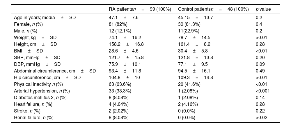
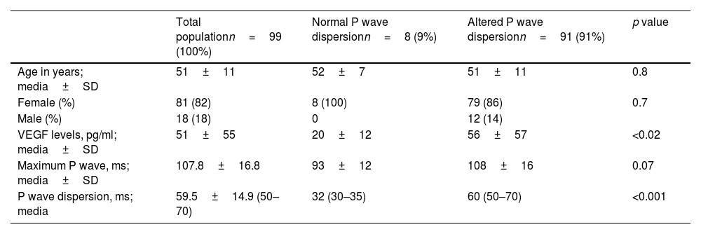
ResultsA total of 99 patients with RA, and 48 control subjects were evaluated. The PWD was 25.3±4.9ms in the control group vs. 57±14.9ms (p<0.0001) in the RA group. No patient in the control group had altered PWD, while 94 (95%) patients in the RA group presented it (p<0.0001). The value of VEGF in the control group was 15.2±15.1pg/ml vs 51.1±55.5pg/ml (p<0.001) in RA. The value of VEGF in RA without altered PWD was 20±12pg/ml vs 56±57pg/ml in RA with altered PWD (p<0.02). An elevated VEGF value had a specificity of 80%, and a positive predictive accuracy of 95% in predicting altered PWD in RA.
ConclusionsThis study establishes for the first time that RA patients who possess significantly higher serum levels of VEGF have an altered PWD. The presence of an elevated VEGF serum value has a high specificity, and high positive predictive accuracy of the existence of altered PWD in RA.
Los niveles séricos del factor de crecimiento endotelial vascular (VEGF) se correlacionan con alteraciones estructurales en la artritis reumatoide (AR). Dado que la dispersión de la onda P (PWD) se asocia con cambios fibróticos relacionados con la isquemia auricular, se concibió que puede haber una correlación entre la PWD alterada y el aumento de los niveles de VEGF en la AR.
MétodosEn este estudio observacional prospectivo, evaluamos a pacientes con AR y los comparamos con sujetos de control. Se consideró la PWD como la diferencia entre la duración máxima y mínima de la onda P. Se consideró la PWD alterada aquella que tenía una dispersión ≥ 38ms. Las mediciones de los niveles séricos de VEGF se realizaron utilizando kits ELISA de medición inmunoabsorvente de enzima-ligando.
ResultadosSe evaluaron un total de 99 pacientes con AR y 48 sujetos controles. La PWD fue de 25,3±4,9ms en el grupo de control frente a 57±14,9ms (P <0,0001) en el grupo de AR. Ningún paciente del grupo control presentó PWD alterada, mientras que 94 (95%) pacientes del grupo AR la presentaron (P<0,0001). El valor de VEGF en el grupo control fue de 15,2±15,1 pg/ml frente a 51,1±55,5 pg/ml en la AR (P <0,001). El valor de VEGF en la AR sin PWD alterada fue de 20±12 pg/ml frente a 56±57 pg/ml en la AR con PWD alterada (P <0,02). Un valor elevado de VEGF tuvo una especificidad del 80% y un valor predictivo positivo del 95% para predecir la PWD alterada en la AR.
ConclusionesEste estudio establece por primera vez que los pacientes con AR que poseen niveles séricos significativamente más altos de VEGF tienen una PWD alterada. La presencia de un valor sérico elevado de VEGF tiene una alta especificidad y un alto valor predictivo positivo de la existencia de PWD alterada en la AR.


