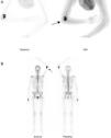
We present the case of a 36 year old female, monitored for painful, progressive tumour in the anteromedial surface of the left elbow and functional deficit, of 2-month onset, which was resistant to analgesic treatment. She referred to a previous history of trauma, and had been diagnosed with fibrillar muscle rupture. Due to the persistence of symptoms, an out-patient MRI was performed which resulted in suspicion of soft tissue sarcoma and the patient was admitted to hospital for further study. Laboratory tests and a CT scan of the chest and abdomen were negative. A two phase bone scan (Fig. 1A and B), showed a heterogeneous uptake area in the anterior region of the left elbow with no relevant findings at any other level. Differential diagnosis was suggested as sarcoma of the soft tissue, chondrosarcoma and osteochondroma with malignant degeneration. It was finally decided that ultrasound-guided percutaneous biopsy be performed, the result of which was mesenchymal tumour without signs of malignancy and final diagnosis of myositis ossificans was made. Rest, anti-inflammatory treatment and physiotherapy were recommended as treatment. Three months later the patient showed clinical signs of improvement and a reduction in pain, with complete flexion and extension at 120°. Plain radiological control (Fig. 2A and B) and noncontrast CT imaging (Fig. 3) revealed a calcified mass, compatible with the anatomopathological diagnosis.
Confined myositis ossificans is a rare entity of uncertain aetiopathogenesis, characterised by a cellular metaplasia of connective tissue induced by trauma.1–3 Examination highlights a hardened tumour which is indistinguishable from other tumour lesions.1 Its main problem arises from generally being confused with malignant (and particularly soft tissue sarcoma) and infectious (osteomyelitis) processes.1,2 It is important to be familiar with the morphological–functional characteristics of this lesion, which evolve at different stages, the acute phase producing an inflammatory reaction with a clinical and radiological pattern that is difficult to distinguish from aggressive conditions.2 The creation of an exhaustive medical history is essential (possible trauma, repeated aggressions, burns, prolonged immobilisation, traumatic paralysis) and an anatomopathologic study to rule out myositis ossificans which is a benign pathology treated in a conservative manner.1–4
Authorship- 1.
Manuscript conception and design: Elena Espinosa Muñoz.
- 2.
Data collection: Elena Espinosa Muñoz.
- 3.
Data analysis and interpretation: Elena Espinosa Muñoz, Diego Ramírez Ocaña, Ana María Martín García and Carmen Puentes Zarzuela.
- 4.
Manuscript redaction, review, and approval: Elena Espinosa Muñoz, Diego Ramírez Ocaña, Ana María Martín García and Carmen Puentes Zarzuela.
The authors declare that for this research no experimentation has been carried out on human beings or animals.
Data confidentialityThe authors declare that no patient data appear in this article.
Right to privacy and informed consentThe authors declare that no patient data appear in this article.
Conflict of InterestsThe authors have no conflicts of interest to declare.
Please cite this article as: Espinosa Muñoz E, Ramírez Ocaña D, Martín García AM, Puentes Zarzuela C. Miositis osificante circunscrita en codo simulando un sarcoma de partes blandas: hallazgos clínico-radiológicos similares. Reumatol Clin. 2019;15:e57–e59.


![Two phase bone scan after the injection of 814MBq of 99mTc-hydroxy-biphosphonate. Static images after 10min (tissue phase [A]) and total body after 2h (bone phase [B]), where heterogeneous uptake may be observed of the radiotracer in anterior region of the left elbow (arrows). Two phase bone scan after the injection of 814MBq of 99mTc-hydroxy-biphosphonate. Static images after 10min (tissue phase [A]) and total body after 2h (bone phase [B]), where heterogeneous uptake may be observed of the radiotracer in anterior region of the left elbow (arrows).](https://static.elsevier.es/multimedia/21735743/0000001500000005/v1_201910260908/S2173574318301217/v1_201910260908/en/main.assets/thumbnail/gr1.jpeg?xkr=ue/ImdikoIMrsJoerZ+w937trqSwLGgTrQM2QjUSRyU=)


