Noonan's syndrome is an autosomal dominant genetic disorder with high phenotypic variability, characterized mainly by facial dysmorphism, congenital heart disease and short stature. We describe the case of a male patient diagnosed with Noonan's syndrome and peripheral spondyloarthritis, a previously undescribed association in the literature.
El síndrome de Noonan es un síndrome genético autosómico dominante que presenta una gran variabilidad fenotípica, caracterizado principalmente por dimorfismo facial, cardiopatía congénita y talla baja. Describimos el caso de un paciente de sexo masculino con síndrome de Noonan y espondiloartritis periférica, asociación no descrita en la literatura hasta el momento.
Noonan's syndrome (NS) is an autosomal dominant genetic syndrome characterized by short stature, cardiac abnormalities, a short neck, chest deformity, a characteristic phenotype with hypertelorism and mental retardation.1 Its incidence so far has been estimated at 1:1000 to 1:2500 live births.2
The clinical characteristics of patients with NS are a triangular face with a broad forehead, hypertelorism, epicanthus, ptosis, a depressed nasal bridge, micrognathia, small ears, short neck and cardiac abnormalities.3,4 Among the musculoskeletal disorders, the most common chest deformities are pectus carinatum and/or pectus excavatum, observed in 70% of cases. In addition there is ulnar valgus clynobrachydactylia, scoliosis/kyphosis, radioulnar synostosis, hyperextensibility and dental malocclusion.5
Other clinical findings are usually bilateral cryptorchidism, which occurs in 60% of male patients, learning and language disorders, attention deficit and depression in 23% of cases.6
NS diagnosis is made with clinical findings, according to the criteria formulated by van der Burgt in 1997 and published in 2007, described in Table 1.7 Treatment is based on the clinical manifestations.8
Van der Burgt Criteria for the Clinical Diagnosis of Noonan's Syndrome.
| Characteristics | Major criteria | Minor criteria | Patient criteria |
|---|---|---|---|
| Facial | Typical face | Suggestive face | Typical face |
| Cardiac | Pulmonary valve stenosisHypertrophic cardiomyopathy and/or ECG changes | Other cardiac abnormalities | Intraventricular communication which was treated surgicallyAt the time, no cardiac abnormalities |
| Height | <3rd percentile | <10th percentile | <3rd percentile |
| Thoracic | Pectus carinatum and/or pectus excavatum | Width | Pectus carinatum |
| Family history | First-degree relative diagnosed with Noonan's syndrome | First degree relative with characteristics suggestive of Noonan's syndrome | First degree relative with characteristics suggestive of Noonan's syndrome |
| Other | Had 3:Mental retardationCryptorchidismLymphatic dysplasia | Had one:Mental retardationCryptorchidismLymphatic dysplasia | Mental retardation |
| Diagnosis of Noonan's syndrome: | |||
| -Two major criteria or one major+2 minor criteria or 3 minor criteria. | |||
| -Typical face with hypertelorism, antimongoloid deviation of palpebral fissures, epicanthus, low-set and rotated ears. | |||
We report the case of a young male patient with a clinical diagnosis of NS, exhibiting peripheral spondyloarthritis. The relevance of this case is the presentation of a genetic disease with spondyloarthritis, entities that generally do not coexist, allowing us to do a literature review and report this as the first clinical association seen in our region.
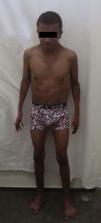
Clinical CaseThe patient is a 23-year-old male with a family history and diagnosis of NS since the age of 2, manifested by cardiac abnormalities (ventricular septal defect diagnosed at 6 months, managed with digoxin, furosemide, antibiotic prophylaxis and surgery until resolved), chest deformity (pectus carinatum), cryptorchidism, bilateral inguinal hernia and surgically corrected phimosis, as well as short stature, a characteristic phenotype with hypertelorism and learning disorders. In 2013 he began a 6 months evolution with ankle, metacarpophalangeal and carpal inflammatory pain, with morning stiffness, and was treated with NSAIDs occasionally with little response; so he was referred to the rheumatology department, with joint pain, bilateral ankle edema and conjunctival hyperemia (Fig. 1).
On physical examination, he had a triangular face with a broad forehead, antimongoloid deviation of palpebral fissures, low-set and rotated ears (Figs. 2 and 3) He presented stunted growth. His height was 1.52cm (<3rd percentile), weight: 38.4kg (<3rd percentile), and he presented pectus carinatum, arthritis of the wrist and fourth metacarpophalangeal of the right hand, second and third proximal interphalangeal joint and distally on the left hand, knee as well as the right knee and ankle, with left anserine bursitis and enthesitis of the left Achilles tendon with bilateral plantar fasciitis. The modified Schober test was reduced (2cm), thoracic expansibility 4cm, lateral lumbar flexion 9cm. Occiput–wall distance was 0cm, tragus–wall 11cm and finger–floor distance of 37cm. The patient had no skin, cardiac or genitourinary abnormalities.
Laboratory tests showed: Hb 13.7mg/dl, Hct 40.8%, WBC: 11,600/mm3; platelets: 456,000/mm3; neutrophils 59%, lymphocytes: 21% CRP: 27 3mg/L; GOT: 13U/L; GPT: 24U/L, GGT: 36U/L, urea 20mg/dl, creatinine 0.8mg/dl, rheumatoid factor: negative, HLA-B27: positive.
X rays of the knees and hands were performed and showed decreased joint space and osteopenia. Magnetic resonance imaging of the lumbosacral spine and hip showed normal vertebral bodies, spinal canal and interapophyseal and sacroiliac joints, with edema of the sacrum. MRI showed edema of the ankles in the posterior region of the calcaneus, thickening and increased density in the insertion of the left Achilles tendon, partial rupture and associated peritendinitis and retrocalcaneal bursitis (Fig. 4). Bone densitometry revealed a lower bone mass than expected for the patients’ age.
The ophthalmologic evaluation revealed mild episcleritis, bilateral normal visual acuity, and the patient was treated with ophthalmic corticosteroid 2 times/day.
The diagnosis of peripheral spondyloarthritis was made based on the ASAS group classification criteria (arthritis, enthesitis plus HLA-B27 positive).9
The BASDAI scale was applied, which measures disease activity in patients with ankylosing spondylitis, with a value of 4. The patient was treated with naproxen 500mg/12h sulfasalazine 1.5g/day, calcium and vitamin D. After 6 months of treatment the patient had a BASDAI of 2, with improvement of clinical symptoms.
DiscussionNS is an uncommon genetic syndrome and an important differential diagnosis in patients with short stature. Musculoskeletal manifestations commonly seen in these patients are pectus Carinatum type chest deformities, observed in 70% of cases, as well as kyphosis or scoliosis.10
Bertola et al. described, in a study of 31 patients with NS, musculoskeletal disorders in 27% of the population, the most prevalent being spina bifida and scoliosis, findings not observed in our patient.11
Pozo also described cubitus valgus (50%), clynobrachydactylia (30%), radioulnar synostosis (2%), hyperextensible joints (50%), thoracic and spinal scoliosis in 25% of cases.12
None of the studies found arthritis and enthesitis related to the genetic syndrome.
The differential diagnosis of NS includes Turner syndrome, Aarskog syndrome, Klippel-Feil syndrome, fetal alcohol syndrome and primidone embryopathy.13
Our patient was diagnosed clinically at 2 years of age and at the time of consultation had major criteria for NS manifested by a typical face, height <3rd percentile, with a first degree family history suggestive of NS, pectus carinatum and absence of cardiac abnormalities associated with the diagnosis of peripheral spondyloarthritis according to the ASAS group criteria.
During initial treatment the patient showed little response to NSAIDs due to their occasional use, taking them only in case of pain. Once the diagnosis of peripheral spondyloarthritis was made and no renal involvement that limited drug use was seen, we started naproxen 500mg/12h sulfasalazine 1.5g/day, calcium and vitamin D with clinical improvement.
After conducting an extensive review of literature in the PubMed and Cochrane databases, we determined that the association of peripheral spondyloarthritis and NS in a young male patient is a relevant fact which no publication to date has documented.
We can also consider that the coexistence of these 2 diseases is a casual relationship and, according to the literature studied, the genetic alterations of NS do not appear to be associated with HLA-B27, which is important for the rheumatologist to know, in addition to the musculoskeletal disorders associated with this genetic syndrome.
Ethical ResponsibilitiesProtection of people and animalsThe authors declare that no experiments have been performed on humans or animals.
Data Privacy. The authors state that no patient data appear in this article.
Right to privacy and informed consentThe authors state that no patient data appear in this article.
Conflict of InterestThe authors declare no conflict of interest.
FinancingNone.
Please cite this article as: Saldarriaga Rivera LM, Fernandes de Melo E, Damião Araujo P, Araujo Silva Filho N, Delgado Quiroz LA, Rios Gomes Bica BE. Presentación de espondiloartritis periférica en paciente con síndrome de Noonan. Reumatol Clin. 2015;11:112–115.


