Información de la revista
Vol. 11. Núm. 4.
Páginas 255-257 (julio - agosto 2015)
Compartir
Compartir
Descargar PDF
Más opciones de artículo
Vol. 11. Núm. 4.
Páginas 255-257 (julio - agosto 2015)
Images in Clinical Rheumatology
Acceso a texto completo
Hypertrophic osteoarthropathy as a complication of pulmonary tuberculosis
Osteoartropatía hipertrófica como complicación de la tuberculosis pulmonar
Visitas
6055
Muñoz Carlos Horacio, Vásquez Gloria María, González Luis Alonso
Autor para correspondencia
lagnvvn68@gmail.com

Corresponding author.
Division of Rheumatology, School of Medicine, Universidad de Antioquia, Hospital Universitario San Vicente Fundación, Medellín, Colombia
Este artículo ha recibido
Información del artículo
Texto completo
Bibliografía
Descargar PDF
Estadísticas
Figuras (3)
Mostrar másMostrar menos
Texto completo

The patient is a 17-year-old man with a history of pulmonary tuberculosis treated for 6 months with the standard regimen [isoniazid, rifampin, ethambutol, and pyrazinamide for 2 months (intensive phase) followed by isoniazid and rifampin for 4 months (continuation phase)] with complete adherence and whose sputum smears at the end of his treatment were negative for acid-fast bacillus. Six months later, he presented to the emergency room complaining of pain and swelling of the ankles, knees and wrists for four weeks prior to admission with no constitutional (fever, night sweats, fatigue, weight loss) and respiratory symptoms. On physical examination, finger clubbing was evident and confirmed by observation of the Schamroth's sign (Fig. 1).

Fig. 1.

Digital clubbing. (A) Clubbed appearance of distal phalanges. (B) Positive Schamroth's sign: the normal diamond-shaped window created by placing the dorsal surfaces of terminal phalanges on opposing fingers together is obliterated in clubbing.

(0.2MB).

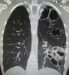
Chest computer tomography showed cystic bronchiectasis and cavitations in the left lower and upper lobes (Fig. 2). X-rays of wrists and legs showed periosteal reaction along the metacarpals, distal radius, ulna, tibia and fibula (Fig. 3) but not acro-osteolysis of the distal phalanges of the hands and feet. Human immunodeficiency virus antibody test was negative. Pharmacologic therapy with nonsteroidal anti-inflammatory drugs was effective in relieving pain from hypertrophic osteoarthropathy (HOA) during his hospital stay and after 3 months of follow-up.

Fig. 2.

Chest computer tomography scan showing cystic bronchiectasis and cavitation in the left lower and upper lobes.

(0.22MB).
Fig. 3.

Radiographs of wrists (A and B) and legs (C) showing periostosis (arrows) at metacarpals, distal ends of the radius, ulna, tibia and fibula.

(0.32MB).
Discussion

HOA is a syndrome characterized by digital clubbing, periostosis of tubular bones and noninflammatory arthritis of large joints.1,2 Digital clubbing is characterized by an increase of the normal 160-degree angle between the nail bed and the proximal nail-fold (Lovibond's angle). In clubbing, this angle exceeds 180 degrees, while in pseudoclubbing it is preserved. Contrary to digital clubbing, finger involvement is asymmetrical in the majority of the reported cases of pseudoclubbing, and acro-osteolysis instead of an overgrowth of the phalangeal tufts seen in clubbing, is the typical radiographic finding in pseudoclubbing.3

HOA is classified as either primary (pachydermoperiostosis) or secondary to pulmonary, cardiac, liver or gastrointestinal diseases characterized by arteriovenous shunt.1,2 Pulmonary malignancies are the most common disorders associated with HOA (80%). Chronic infections associated with cystic fibrosis, subacute bacterial endocarditis, acquired immunodeficiency syndrome and pulmonary tuberculosis are also associated with HOA.4 Our case illustrates the HOA as a rare late complication of pulmonary tuberculosis.

The pathogenesis of HOA is unknown, although an impaired lung function due to the exclusion of this organ from part of the circulation is common in most diseases associated with HOA. It has been suggested that in hypoxic conditions, megakaryocytes bypass the pulmonary circulation through arteriovenous shunts and directly reach the distal sites in the systemic circulation and interact with endothelial cells, releasing growth factors, such as vascular endothelial growth factor (VEGF), resulting in angiogenesis and thus digital clubbing.1,5•8 VEGF is a platelet-derived factor induced by hypoxia that produces vascular hyperplasia, new bone formation, edema, and fibroblast/osteoblast proliferation, which are histological alterations of HOA.6•8

Although the association of HOA with pulmonary tuberculosis has been rarely reported,9•12 physicians should consider HOA as a complication of non-neoplastic diseases, such as pulmonary tuberculosis, an endemic infection of the world's population.

Conflicts of interest

The authors declare no conflicts of interest.

References
[1]
M. Martínez-Lavín, C. Pineda.
Digital clubbing and hypertrophic osteoarthropathy.
Rheumatology, 5th ed., pp. 1701-1705
[2]
C. Pineda, M. Martínez-Lavín.
Hypertrophic osteoarthropathy: what a rheumatologist should know about this uncommon condition.
Rheum Dis Clin North Am, 39 (2013), pp. 383-400
[3]
M.B. Santiago, I. Lima, A.C. Feitosa, S. Braz Ade, L.G. Miranda.
Pseudoclubbing: is it different from clubbing?.
Semin Arthritis Rheum, 38 (2009), pp. 452-457
[4]
Q. Yao, R.D. Altman, E. Brahn.
Periostitis and hypertrophic pulmonary osteoarthropathy: report of 2 cases and review of the literature.
Semin Arthritis Rheum, 38 (2009), pp. 458-466
[5]
C.J. Dickinson, J.F. Martin.
Megakaryocytes and platelet clumps as the cause of finger clubbing.
Lancet, 2 (1987), pp. 1434-1435
[6]
S. Atkinson, S.B. Fox.
Vascular endothelial growth factor (VEGF)-A and platelet-derived growth factor (PDGF) play a central role in the pathogenesis of digital clubbing.
J Pathol, 203 (2004), pp. 721-728
[7]
M. Martinez-Lavin.
Exploring the cause of the most ancient clinical sign of medicine: finger clubbing.
Semin Arthritis Rheum, 36 (2007), pp. 380-385
[8]
F. Olán, M. Portela, C. Navarro, M. Gaxiola, L.H. Silveira, V. Ruiz, et al.
Circulating vascular endothelial growth factor concentrations in a case of pulmonary hypertrophic osteoarthropathy. Correlation with disease activity.
J Rheumatol, 31 (2004), pp. 614-616
[9]
P. Kelly, P. Manning, P. Corcoran, L. Clancy.
Hypertrophic osteoarthropathy in association with pulmonary tuberculosis.
Chest, 99 (1991), pp. 769-770
[10]
I. Aissa, A. Messadi, S. Boudaya, F. El Mezni, T. Kilani, H. Ghèc)dira.
Hypertrophic osteoarthropathy associated with pulmonary tuberculosis.
Rev Pneumol Clin, 67 (2011), pp. 101-104
[11]
J.G. Webb, P. Thomas.
Hypertrophic osteoarthropathy in pulmonary tuberculosis.
Tubercle, 67 (1986), pp. 225-228
[12]
P.V. Barber, R. Lechler.
Hypertrophic osteoarthropathy: two unusual causes.
Postgrad Med J, 59 (1983), pp. 254-255
Copyright © 2014. Elsevier España, S.L.U.. All rights reserved
Descargar PDF
Idiomas
Reumatología Clínica
Opciones de artículo
Herramientas
es en

¿Es usted profesional sanitario apto para prescribir o dispensar medicamentos?

Are you a health professional able to prescribe or dispense drugs?