array:24 [
  "pii" => "S2173574312000160"
  "issn" => "21735743"
  "doi" => "10.1016/j.reumae.2011.10.007"
  "estado" => "S300"
  "fechaPublicacion" => "2012-09-01"
  "aid" => "381"
  "copyright" => "Elsevier España, S.L.. All rights reserved"
  "copyrightAnyo" => "2011"
  "documento" => "article"
  "crossmark" => 0
  "subdocumento" => "sco"
  "cita" => "Reumatol Clin. 2012;8:294-7"
  "abierto" => array:3 [
    "ES" => false
    "ES2" => false
    "LATM" => false
  ]
  "gratuito" => false
  "lecturas" => array:2 [
    "total" => 11199
    "formatos" => array:3 [
      "EPUB" => 68
      "HTML" => 10162
      "PDF" => 969
    ]
  ]
  "Traduccion" => array:1 [
    "es" => array:19 [
      "pii" => "S1699258X11003184"
      "issn" => "1699258X"
      "doi" => "10.1016/j.reuma.2011.10.010"
      "estado" => "S300"
      "fechaPublicacion" => "2012-09-01"
      "aid" => "381"
      "copyright" => "Elsevier España, S.L."
      "documento" => "article"
      "crossmark" => 0
      "subdocumento" => "sco"
      "cita" => "Reumatol Clin. 2012;8:294-7"
      "abierto" => array:3 [
        "ES" => true
        "ES2" => true
        "LATM" => true
      ]
      "gratuito" => true
      "lecturas" => array:2 [
        "total" => 21090
        "formatos" => array:3 [
          "EPUB" => 162
          "HTML" => 18718
          "PDF" => 2210
        ]
      ]
      "es" => array:11 [
        "idiomaDefecto" => true
        "cabecera" => "<span class="elsevierStyleTextfn">Reumatolog&#237;a cl&#237;nica en im&#225;genes</span>"
        "titulo" => "Tofo tibial intra&#243;seo en gota cr&#243;nica"
        "tienePdf" => "es"
        "tieneTextoCompleto" => "es"
        "paginas" => array:1 [
          0 => array:2 [
            "paginaInicial" => "294"
            "paginaFinal" => "297"
          ]
        ]
        "titulosAlternativos" => array:1 [
          "en" => array:1 [
            "titulo" => "Intrabony tibial tophi in chronic gout"
          ]
        ]
        "contieneTextoCompleto" => array:1 [
          "es" => true
        ]
        "contienePdf" => array:1 [
          "es" => true
        ]
        "resumenGrafico" => array:2 [
          "original" => 0
          "multimedia" => array:7 [
            "identificador" => "fig0010"
            "etiqueta" => "Figura 2"
            "tipo" => "MULTIMEDIAFIGURA"
            "mostrarFloat" => true
            "mostrarDisplay" => false
            "figura" => array:1 [
              0 => array:4 [
                "imagen" => "gr2.jpeg"
                "Alto" => 1189
                "Ancho" => 950
                "Tamanyo" => 69417
              ]
            ]
            "descripcion" => array:1 [
              "es" => "<p id="spar0010" class="elsevierStyleSimplePara elsevierViewall">Radiograf&#237;a de rodilla izquierda anteroposterior&#58; se objetivan lesiones l&#237;ticas de bordes bien delimitados en ep&#237;fisis tibial y cambios degenerativos incipientes femorotibiales&#46;</p>"
            ]
          ]
        ]
        "autores" => array:1 [
          0 => array:2 [
            "autoresLista" => "Crist&#243;bal M&#46; Rodr&#237;guez Leal, Raquel Almod&#243;var, Pedro Zarco, Ram&#243;n Mazzuchelli, Francisco Javier Quir&#243;s"
            "autores" => array:5 [
              0 => array:2 [
                "nombre" => "Crist&#243;bal M&#46;"
                "apellidos" => "Rodr&#237;guez Leal"
              ]
              1 => array:2 [
                "nombre" => "Raquel"
                "apellidos" => "Almod&#243;var"
              ]
              2 => array:2 [
                "nombre" => "Pedro"
                "apellidos" => "Zarco"
              ]
              3 => array:2 [
                "nombre" => "Ram&#243;n"
                "apellidos" => "Mazzuchelli"
              ]
              4 => array:2 [
                "nombre" => "Francisco Javier"
                "apellidos" => "Quir&#243;s"
              ]
            ]
          ]
        ]
      ]
      "idiomaDefecto" => "es"
      "Traduccion" => array:1 [
        "en" => array:9 [
          "pii" => "S2173574312000160"
          "doi" => "10.1016/j.reumae.2011.10.007"
          "estado" => "S300"
          "subdocumento" => ""
          "abierto" => array:3 [
            "ES" => false
            "ES2" => false
            "LATM" => false
          ]
          "gratuito" => false
          "lecturas" => array:1 [
            "total" => 0
          ]
          "idiomaDefecto" => "en"
          "EPUB" => "https://multimedia.elsevier.es/PublicationsMultimediaV1/item/epub/S2173574312000160?idApp=UINPBA00004M"
        ]
      ]
      "EPUB" => "https://multimedia.elsevier.es/PublicationsMultimediaV1/item/epub/S1699258X11003184?idApp=UINPBA00004M"
      "url" => "/1699258X/0000000800000005/v1_201305061926/S1699258X11003184/v1_201305061926/es/main.assets"
    ]
  ]
  "itemSiguiente" => array:19 [
    "pii" => "S2173574312000779"
    "issn" => "21735743"
    "doi" => "10.1016/j.reumae.2012.06.003"
    "estado" => "S300"
    "fechaPublicacion" => "2012-09-01"
    "aid" => "420"
    "copyright" => "Elsevier Espa&#241;a&#44; S&#46;L&#46;"
    "documento" => "simple-article"
    "crossmark" => 0
    "subdocumento" => "cor"
    "cita" => "Reumatol Clin. 2012;8:298-9"
    "abierto" => array:3 [
      "ES" => false
      "ES2" => false
      "LATM" => false
    ]
    "gratuito" => false
    "lecturas" => array:2 [
      "total" => 3442
      "formatos" => array:3 [
        "EPUB" => 58
        "HTML" => 2777
        "PDF" => 607
      ]
    ]
    "en" => array:10 [
      "idiomaDefecto" => true
      "cabecera" => "<span class="elsevierStyleTextfn">Letter to the Editor</span>"
      "titulo" => "Transverse Myelitis and Bilateral Optic Neuritis in a Patient With Systemic Lupus Erythematosus"
      "tienePdf" => "en"
      "tieneTextoCompleto" => "en"
      "paginas" => array:1 [
        0 => array:2 [
          "paginaInicial" => "298"
          "paginaFinal" => "299"
        ]
      ]
      "titulosAlternativos" => array:1 [
        "es" => array:1 [
          "titulo" => "Mielitis transversa y neuritis &#243;ptica bilateral en una paciente con lupus eritematoso sist&#233;mico"
        ]
      ]
      "contieneTextoCompleto" => array:1 [
        "en" => true
      ]
      "contienePdf" => array:1 [
        "en" => true
      ]
      "autores" => array:1 [
        0 => array:2 [
          "autoresLista" => "I&#241;aki Hernando Rubio, Joaqu&#237;n Belzunegui Otano, Olga M&#225;iz Alonso, Bel&#233;n &#193;lvarez Rodr&#237;guez"
          "autores" => array:4 [
            0 => array:2 [
              "nombre" => "I&#241;aki"
              "apellidos" => "Hernando Rubio"
            ]
            1 => array:2 [
              "nombre" => "Joaqu&#237;n"
              "apellidos" => "Belzunegui Otano"
            ]
            2 => array:2 [
              "nombre" => "Olga"
              "apellidos" => "M&#225;iz Alonso"
            ]
            3 => array:2 [
              "nombre" => "Bel&#233;n"
              "apellidos" => "&#193;lvarez Rodr&#237;guez"
            ]
          ]
        ]
      ]
    ]
    "idiomaDefecto" => "en"
    "Traduccion" => array:1 [
      "es" => array:9 [
        "pii" => "S1699258X12000526"
        "doi" => "10.1016/j.reuma.2012.01.005"
        "estado" => "S300"
        "subdocumento" => ""
        "abierto" => array:3 [
          "ES" => true
          "ES2" => true
          "LATM" => true
        ]
        "gratuito" => true
        "lecturas" => array:1 [
          "total" => 0
        ]
        "idiomaDefecto" => "es"
        "EPUB" => "https://multimedia.elsevier.es/PublicationsMultimediaV1/item/epub/S1699258X12000526?idApp=UINPBA00004M"
      ]
    ]
    "EPUB" => "https://multimedia.elsevier.es/PublicationsMultimediaV1/item/epub/S2173574312000779?idApp=UINPBA00004M"
    "url" => "/21735743/0000000800000005/v1_201305061645/S2173574312000779/v1_201305061645/en/main.assets"
  ]
  "itemAnterior" => array:19 [
    "pii" => "S2173574312000172"
    "issn" => "21735743"
    "doi" => "10.1016/j.reumae.2011.10.008"
    "estado" => "S300"
    "fechaPublicacion" => "2012-09-01"
    "aid" => "382"
    "copyright" => "Elsevier Espa&#241;a&#44; S&#46;L&#46;"
    "documento" => "article"
    "crossmark" => 0
    "subdocumento" => "sco"
    "cita" => "Reumatol Clin. 2012;8:292-3"
    "abierto" => array:3 [
      "ES" => false
      "ES2" => false
      "LATM" => false
    ]
    "gratuito" => false
    "lecturas" => array:2 [
      "total" => 7761
      "formatos" => array:3 [
        "EPUB" => 57
        "HTML" => 6982
        "PDF" => 722
      ]
    ]
    "en" => array:11 [
      "idiomaDefecto" => true
      "cabecera" => "<span class="elsevierStyleTextfn">Images in Clinical Rheumatology</span>"
      "titulo" => "Tuberculous Spondylodiskitis With Lumbar Tumor"
      "tienePdf" => "en"
      "tieneTextoCompleto" => "en"
      "paginas" => array:1 [
        0 => array:2 [
          "paginaInicial" => "292"
          "paginaFinal" => "293"
        ]
      ]
      "titulosAlternativos" => array:1 [
        "es" => array:1 [
          "titulo" => "Espondilodiscitis tuberculosa con tumoraci&#243;n lumbar"
        ]
      ]
      "contieneTextoCompleto" => array:1 [
        "en" => true
      ]
      "contienePdf" => array:1 [
        "en" => true
      ]
      "resumenGrafico" => array:2 [
        "original" => 0
        "multimedia" => array:7 [
          "identificador" => "fig0015"
          "etiqueta" => "Fig&#46; 3"
          "tipo" => "MULTIMEDIAFIGURA"
          "mostrarFloat" => true
          "mostrarDisplay" => false
          "figura" => array:1 [
            0 => array:4 [
              "imagen" => "gr3.jpeg"
              "Alto" => 1199
              "Ancho" => 998
              "Tamanyo" => 112635
            ]
          ]
          "descripcion" => array:1 [
            "en" => "<p id="spar0015" class="elsevierStyleSimplePara elsevierViewall">L1 and L2 lytic lesion with a sclerotic border&#46;</p>"
          ]
        ]
      ]
      "autores" => array:1 [
        0 => array:2 [
          "autoresLista" => "Mar&#237;a Cristina L&#243;pez-S&#225;nchez, Gabriela Calvo Arrojo, Tom&#225;s Ram&#243;n V&#225;zquez-Rodr&#237;guez"
          "autores" => array:3 [
            0 => array:2 [
              "nombre" => "Mar&#237;a Cristina"
              "apellidos" => "L&#243;pez-S&#225;nchez"
            ]
            1 => array:2 [
              "nombre" => "Gabriela"
              "apellidos" => "Calvo Arrojo"
            ]
            2 => array:2 [
              "nombre" => "Tom&#225;s Ram&#243;n"
              "apellidos" => "V&#225;zquez-Rodr&#237;guez"
            ]
          ]
        ]
      ]
    ]
    "idiomaDefecto" => "en"
    "Traduccion" => array:1 [
      "es" => array:9 [
        "pii" => "S1699258X11003196"
        "doi" => "10.1016/j.reuma.2011.10.011"
        "estado" => "S300"
        "subdocumento" => ""
        "abierto" => array:3 [
          "ES" => true
          "ES2" => true
          "LATM" => true
        ]
        "gratuito" => true
        "lecturas" => array:1 [
          "total" => 0
        ]
        "idiomaDefecto" => "es"
        "EPUB" => "https://multimedia.elsevier.es/PublicationsMultimediaV1/item/epub/S1699258X11003196?idApp=UINPBA00004M"
      ]
    ]
    "EPUB" => "https://multimedia.elsevier.es/PublicationsMultimediaV1/item/epub/S2173574312000172?idApp=UINPBA00004M"
    "url" => "/21735743/0000000800000005/v1_201305061645/S2173574312000172/v1_201305061645/en/main.assets"
  ]
  "en" => array:15 [
    "idiomaDefecto" => true
    "cabecera" => "<span class="elsevierStyleTextfn">Images in Clinical Rheumatology</span>"
    "titulo" => "Intrabony Tibial Tophi in Chronic Gout"
    "tieneTextoCompleto" => true
    "paginas" => array:1 [
      0 => array:2 [
        "paginaInicial" => "294"
        "paginaFinal" => "297"
      ]
    ]
    "autores" => array:1 [
      0 => array:4 [
        "autoresLista" => "Crist&#243;bal M&#46; Rodr&#237;guez Leal, Raquel Almod&#243;var, Pedro Zarco, Ram&#243;n Mazzuchelli, Francisco Javier Quir&#243;s"
        "autores" => array:5 [
          0 => array:3 [
            "nombre" => "Crist&#243;bal M&#46;"
            "apellidos" => "Rodr&#237;guez Leal"
            "referencia" => array:1 [
              0 => array:2 [
                "etiqueta" => "<span class="elsevierStyleSup">a</span>"
                "identificador" => "aff0005"
              ]
            ]
          ]
          1 => array:4 [
            "nombre" => "Raquel"
            "apellidos" => "Almod&#243;var"
            "email" => array:1 [
              0 => "ralmodovar&#64;fhalcorcon&#46;es"
            ]
            "referencia" => array:2 [
              0 => array:2 [
                "etiqueta" => "<span class="elsevierStyleSup">b</span>"
                "identificador" => "aff0010"
              ]
              1 => array:2 [
                "etiqueta" => "<span class="elsevierStyleSup">¿</span>"
                "identificador" => "cor0005"
              ]
            ]
          ]
          2 => array:3 [
            "nombre" => "Pedro"
            "apellidos" => "Zarco"
            "referencia" => array:1 [
              0 => array:2 [
                "etiqueta" => "<span class="elsevierStyleSup">b</span>"
                "identificador" => "aff0010"
              ]
            ]
          ]
          3 => array:3 [
            "nombre" => "Ram&#243;n"
            "apellidos" => "Mazzuchelli"
            "referencia" => array:1 [
              0 => array:2 [
                "etiqueta" => "<span class="elsevierStyleSup">b</span>"
                "identificador" => "aff0010"
              ]
            ]
          ]
          4 => array:3 [
            "nombre" => "Francisco Javier"
            "apellidos" => "Quir&#243;s"
            "referencia" => array:1 [
              0 => array:2 [
                "etiqueta" => "<span class="elsevierStyleSup">b</span>"
                "identificador" => "aff0010"
              ]
            ]
          ]
        ]
        "afiliaciones" => array:2 [
          0 => array:3 [
            "entidad" => "Unidad de Medicina Interna&#44; Hospital Universitario Fundaci&#243;n Alcorc&#243;n&#44; Madrid&#44; Spain"
            "etiqueta" => "<span class="elsevierStyleSup">a</span>"
            "identificador" => "aff0005"
          ]
          1 => array:3 [
            "entidad" => "Unidad de Reumatolog&#237;a&#44; Hospital Universitario Fundaci&#243;n Alcorc&#243;n&#44; Madrid&#44; Spain"
            "etiqueta" => "<span class="elsevierStyleSup">b</span>"
            "identificador" => "aff0010"
          ]
        ]
        "correspondencia" => array:1 [
          0 => array:3 [
            "identificador" => "cor0005"
            "etiqueta" => "&#8270;"
            "correspondencia" => "Corresponding author&#46;"
          ]
        ]
      ]
    ]
    "titulosAlternativos" => array:1 [
      "es" => array:1 [
        "titulo" => "Tofo tibial intra&#243;seo en gota cr&#243;nica"
      ]
    ]
    "resumenGrafico" => array:2 [
      "original" => 0
      "multimedia" => array:7 [
        "identificador" => "fig0015"
        "etiqueta" => "Fig&#46; 3"
        "tipo" => "MULTIMEDIAFIGURA"
        "mostrarFloat" => true
        "mostrarDisplay" => false
        "figura" => array:1 [
          0 => array:4 [
            "imagen" => "gr3a.jpeg"
            "Alto" => 917
            "Ancho" => 944
            "Tamanyo" => 128537
          ]
        ]
        "descripcion" => array:1 [
          "en" => "<p id="spar0015" class="elsevierStyleSimplePara elsevierViewall">&#40;A&#41; Left knee coronal T1MRI&#46; It shows a diffuse hypointense synovial proliferation with bone erosion of the posterolateral margin of the femoral condyle &#40;blue arrow&#41; and intercondylar notch&#46; A hypointense lytic lesion is seen in the proximal tibial epiphysis with a maximum diameter of 5<span class="elsevierStyleHsp" style=""></span>cm &#40;red arrows&#41;&#46; &#40;B&#41; Sagittal T2&#46; There is an increased signal heterogeneity at the level of the tibial epiphyseal lytic lesion &#40;red arrows&#41;&#44; along with a large Baker&#39;s cyst located behind the medial gastrocnemius muscle &#40;green arrows&#41;&#44; with areas of high and low signal for synovial hypertrophy&#46; &#40;C&#41; Sagittal T1-Gd&#46; After administration of gadolinium&#44; enhancement shows the level of peripheral tibial lytic lesion &#40;red arrows&#41; and Baker&#39;s cyst &#40;green arrows&#41;&#46; &#40;For interpretation of the references to color in this figure legend&#44; the reader is referred to the web version of the article&#46;&#41;</p>"
        ]
      ]
    ]
    "textoCompleto" => "<span class="elsevierStyleSections"><span id="sec0005" class="elsevierStyleSection elsevierViewall"><span class="elsevierStyleSectionTitle">Case Report</span><p id="par0005" class="elsevierStylePara elsevierViewall">We present the case of a 49-year-old male with a history of hyperlipidemia and significant alcohol intake&#46; In May 2004&#44; during an outpatient visit&#44; he referred 10 years of recurrent episodes of monoarthritis lasting three days&#44; successively affecting either the left knee&#44; left tarsus&#44; elbow&#44; left or bilateral first metatarsophalangeal joints&#44; with no other accompanying symptoms&#46; Physical examination showed signs of a left knee effusion&#44; restricting flexion to 90&#176; and tophi on the left olecranon&#46;</p><p id="par0010" class="elsevierStylePara elsevierViewall">Laboratory tests showed uricemia of 9&#46;4<span class="elsevierStyleHsp" style=""></span>mg&#47;dl and GGT 166<span class="elsevierStyleHsp" style=""></span>U&#47;dl&#46; The remaining parameters &#40;CBC&#44; TSH&#44; CPK&#44; rheumatoid factor&#44; HLA B27&#44; anti-CCP antibodies&#44; immunoglobulins&#44; 24<span class="elsevierStyleHsp" style=""></span>h urine uric acid&#41; were normal&#46; X-rays of the left foot were performed &#40;<a class="elsevierStyleCrossRef" href="#fig0005">Fig&#46; 1</a>&#41;&#44; showing marginal erosions and sclerotic borders of the first metatarsal head&#44; with increased adjacent soft tissue&#46; A left knee radiograph &#40;<a class="elsevierStyleCrossRef" href="#fig0010">Fig&#46; 2</a>&#41; showed lytic lesions with well-defined edges and incipient degenerative changes of the tibial epiphysis at the femorotibial level&#46; We performed arthrocentesis of the left knee&#46; Monosodium urate crystals were found in synovial fluid &#40;SF&#41;&#46; SF culture was negative&#46;</p><elsevierMultimedia ident="fig0005"></elsevierMultimedia><elsevierMultimedia ident="fig0010"></elsevierMultimedia></span><span id="sec0010" class="elsevierStyleSection elsevierViewall"><span class="elsevierStyleSectionTitle">Diagnosis and Evolution</span><p id="par0015" class="elsevierStylePara elsevierViewall">Given the radiological lesions of the left knee&#44; we completed the study with a left knee MRI &#40;<a class="elsevierStyleCrossRef" href="#fig0015">Fig&#46; 3</a>&#41; which showed synovial proliferation which revealed diffuse&#44; hypointense lesions on T1&#44; with bone erosions in the posterolateral margin of the femoral condyle&#46; A large Baker&#39;s cyst with areas of high and low signal on T2 were seen&#44; as well as lytic&#44; hypointense lesion on T1&#44; T2 with a heterogeneous signal level on the proximal tibial epiphysis and a maximum diameter of 5<span class="elsevierStyleHsp" style=""></span>cm that after administration of gadolinium showed enhancement&#44; something characteristic of tophaceous deposits&#46;</p><elsevierMultimedia ident="fig0015"></elsevierMultimedia><p id="par0020" class="elsevierStylePara elsevierViewall">Based on the above&#44; the patient was diagnosed with chronic gouty arthritis with intraosseous and intraarticular tophi&#46; Dietary measures were initiated and treatment begun with allopurinol &#40;300<span class="elsevierStyleHsp" style=""></span>mg&#47;d&#41; and colchicine 1<span class="elsevierStyleHsp" style=""></span>mg daily&#46; Due to the persistence of clinical uricemia of 6&#46;2<span class="elsevierStyleHsp" style=""></span>mg&#47;dl&#44; benzbromarone was added &#40;up to 100<span class="elsevierStyleHsp" style=""></span>mg doses per day&#41;&#44; until the patient was asymptomatic&#46; Current levels of uric acid are 3&#46;6<span class="elsevierStyleHsp" style=""></span>mg&#47;dl and there was a slight decrease of the tibial intraosseous tophus size&#46;</p></span><span id="sec0015" class="elsevierStyleSection elsevierViewall"><span class="elsevierStyleSectionTitle">Discussion</span><p id="par0025" class="elsevierStylePara elsevierViewall">It is estimated that the prevalence of gouty arthritis in the general population is 1&#46;4&#37;&#44;<a class="elsevierStyleCrossRef" href="#bib0005"><span class="elsevierStyleSup">1</span></a> and represents approximately 5&#37; of monoarthritis seen in consultation&#46; The tophus is a nodular mass composed of sodium urate salts and surrounded by a chronic inflammatory reaction with abundant vascularization&#46; It is a late complication of hyperuricemia maintained and present in only a minority of patients&#46; They usually form in juxta-articular regions&#44; although virtually any tissue<a class="elsevierStyleCrossRef" href="#bib0010"><span class="elsevierStyleSup">2</span></a> can present them&#46; It is not uncommon for intraosseous tophi to form in advanced crystal arthropathy&#44; reaching 13&#46;3&#37; in patients in some series&#46;<a class="elsevierStyleCrossRef" href="#bib0015"><span class="elsevierStyleSup">3</span></a> A definitive diagnosis requires histopathological confirmation&#44; but imaging tests may be useful&#46; Plain radiographs can show juxta-articular and intraosseous calcifications and erosions of the articular surface&#44; creating problems of differential diagnosis with avascular<a class="elsevierStyleCrossRef" href="#bib0020"><span class="elsevierStyleSup">4</span></a> necrosis and osteochondroma&#46; The presence of bone erosions is a marker of intrabone<a class="elsevierStyleCrossRef" href="#bib0025"><span class="elsevierStyleSup">5</span></a> tophi&#46; These can be seen as a soft tissue or intraosseous mass&#46; The joint space is generally respected and periosteal new bone formation and subchondral collapse can appear&#46; Is a useful tool to determine the severity of involvement&#44; with standardized methods and a Sharp&#47;Van der Heijde<a class="elsevierStyleCrossRef" href="#bib0030"><span class="elsevierStyleSup">6</span></a> score available for indexing&#46; Ultrasound is a useful tool for its low cost&#44; no radiation&#44; and speed of implementation&#44; also used to guide invasive techniques&#46; It has a high sensitivity for detecting monosodium urate deposits&#44; even in patients with asymptomatic hyperuricemia&#46; It is able to appreciate bone erosions earlier than plain X-rays&#46; On the other hand&#44; it is the only standardized technique for measurement of tophi and can demonstrate subclinical inflammatory activity by capturing Doppler hypervascular areas around the monosodium urate deposits&#46; The typical findings in gout with this technique are&#58; a double contour sign&#44; aggregated punctate echogenic tissue&#44; &#8220;snow storm&#8221; synovial fluid appearance with hyperechogenic areas&#44; and soft tissue edema&#44; in addition to the aforementioned bone erosions and hypervascularity&#46;<a class="elsevierStyleCrossRefs" href="#bib0030"><span class="elsevierStyleSup">6&#8211;8</span></a> Computed tomography &#40;CT&#41; is useful in the recognition of monosodium urate deposits&#46; These deposits have an attenuation of approximately 160 Hounsfield units &#40;HU&#41;&#44; while calcium crystals exhibit an attenuation of 450<span class="elsevierStyleHsp" style=""></span>HU&#44; even bone tissue and calcified tophi&#46;<a class="elsevierStyleCrossRef" href="#bib0040"><span class="elsevierStyleSup">8</span></a> It provides more specific images than ultrasound or MRI&#44; as well as aid in the diagnosis of complications&#44; and intraosseous tophi and bone erosions&#44; and can guide joint aspiration&#46;<a class="elsevierStyleCrossRef" href="#bib0030"><span class="elsevierStyleSup">6</span></a> Magnetic resonance imaging &#40;MRI&#41; is a tool that&#44; while non-specific and with certain limitations&#44; such as suboptimal definition for intraosseous abnormalities&#44; a high cost&#44; and poor availability&#44; is the method of choice for evaluating soft tissue&#44; ligaments&#44; muscles and tissues as well as juxta-articular joint space&#44; and effusions or synovial proliferation&#44; cartilage abnormalities&#44; due to a spatial resolution far superior to other imaging tests&#46; In particular&#44; it has great utility in determining the etiology of functional limitations of unknown causes in the knee joint in patients with gout&#46;<a class="elsevierStyleCrossRefs" href="#bib0010"><span class="elsevierStyleSup">2&#44;6</span></a> Importantly&#44; the synovial membrane is too thin to be assessed with this imaging method&#44; so that it can only be seen when a pathological process enlarges it&#46; The differential diagnosis of synovial thickening raises the possibility of longstanding rheumatoid arthritis&#44; pyrophosphate arthropathy&#44; villonodular synovitis&#44; hemophilic arthropathy&#44; amyloid arthropathy&#44; synovial chondromatosis&#44; and chronic granulomatous processes such as arthritis or fungal infection or tuberculosis&#46;<a class="elsevierStyleCrossRefs" href="#bib0010"><span class="elsevierStyleSup">2&#44;6&#44;9</span></a> Gout is often found presenting a synovial effusion and synovial pannus&#46; The tophi are seen as an amorphous lesion&#44; eccentric and asymmetric&#44; with variable intensity depending on their composition&#58; protein&#44; fibrous crystals&#44; hemosiderin&#44; and calcium&#44; although the latter are rare and found in very old lesions&#46; It is common to observe one of these morphological patterns&#58; amorphous mass&#44; linear crystalline deposits&#44; or cystic lesions in the juxta-articular bursa&#46; Intraosseous tophi are hypointense or of intermediate intensity on T1&#44; heterogeneous-looking from hyper-to hypointense on T2&#44; depending on the amount of calcium in the tophi&#44; with more calcium&#44; less intensity&#46; It usually enhances after contrast administration&#44; which translates hypervascularization and is a predictor of bone erosion&#46; It also often is accompanied by juxta-articular findings such as erosions&#44; edema of soft tissues&#44; bone marrow edema&#44; or extraarticular<a class="elsevierStyleCrossRef" href="#bib0010"><span class="elsevierStyleSup">2</span></a> tophi&#46;<a class="elsevierStyleCrossRefs" href="#bib0015"><span class="elsevierStyleSup">3&#44;6&#44;9&#8211;11</span></a> Isolated tophi may pose problems in differential diagnosis with tumors such as fibroblastic tumors and benign xantofibroma&#44; but it is rare to find surrounding edema in these&#46;<a class="elsevierStyleCrossRef" href="#bib0050"><span class="elsevierStyleSup">10</span></a></p><p id="par0030" class="elsevierStylePara elsevierViewall">In short&#44; chronic tophaceous gout presents a constellation of clinical and radiological findings which are important to know for a correct differential diagnosis and further treatment&#46; Although MRI is not a specific test for the diagnosis of gout&#44; it does allow early detection of intraosseous and intraarticular tophi and bone erosions in patients with gout&#44; helping us to recognize this entity and avoid a bone biopsy in selected patients&#46; Thus&#44; MRI can play a special role in the identification of clinical complications and monitoring response to treatment&#46;</p></span><span id="sec0020" class="elsevierStyleSection elsevierViewall"><span class="elsevierStyleSectionTitle">Ethical disclosures</span><p id="par0035" class="elsevierStylePara elsevierViewall"><span class="elsevierStyleBold">Protection of human and animal subjects&#46;</span> The authors declare that no experiments were performed on humans or animals for this investigation&#46;</p><p id="par0040" class="elsevierStylePara elsevierViewall"><span class="elsevierStyleBold">Confidentiality of Data&#46;</span> The authors declare that they have followed the protocols of their work centre on the publication of patient data and that all the patients included in the study have received sufficient information and have given their informed consent in writing to participate in that study&#46;</p><p id="par0045" class="elsevierStylePara elsevierViewall"><span class="elsevierStyleBold">Right to privacy and informed consent&#46;</span> The authors have obtained the informed consent of the patients and &#47;or subjects mentioned in the article&#46; The author for correspondence is in possession of this document&#46;</p></span></span>"
    "textoCompletoSecciones" => array:1 [
      "secciones" => array:5 [
        0 => array:2 [
          "identificador" => "sec0005"
          "titulo" => "Case Report"
        ]
        1 => array:2 [
          "identificador" => "sec0010"
          "titulo" => "Diagnosis and Evolution"
        ]
        2 => array:2 [
          "identificador" => "sec0015"
          "titulo" => "Discussion"
        ]
        3 => array:2 [
          "identificador" => "sec0020"
          "titulo" => "Ethical disclosures"
        ]
        4 => array:1 [
          "titulo" => "References"
        ]
      ]
    ]
    "pdfFichero" => "main.pdf"
    "tienePdf" => true
    "NotaPie" => array:1 [
      0 => array:2 [
        "etiqueta" => "&#9734;"
        "nota" => "<p class="elsevierStyleNotepara">Please cite this article as&#58; Rodr&#237;guez Leal CM&#44; et al&#46; Tofo tibial intra&#243;seo en gota cr&#243;nica&#46; Reumatol Clin&#46; 2012&#46; <span class="elsevierStyleInterRef" href="doi:10.1016/j.reuma.2011.10.010">doi&#58;10&#46;1016&#47;j&#46;reuma&#46;2011&#46;10&#46;010</span>&#46;</p>"
      ]
    ]
    "multimedia" => array:4 [
      0 => array:7 [
        "identificador" => "fig0005"
        "etiqueta" => "Fig&#46; 1"
        "tipo" => "MULTIMEDIAFIGURA"
        "mostrarFloat" => true
        "mostrarDisplay" => false
        "figura" => array:1 [
          0 => array:4 [
            "imagen" => "gr1.jpeg"
            "Alto" => 1887
            "Ancho" => 1501
            "Tamanyo" => 155504
          ]
        ]
        "descripcion" => array:1 [
          "en" => "<p id="spar0005" class="elsevierStyleSimplePara elsevierViewall">Anteroposterior and oblique X-ray of left foot&#58; well demarcated marginal erosions are observed on the first metatarsal head&#44; with increased adjacent soft tissue&#46; The joint space and the density of the rest of the subchondral bone is preserved&#46;</p>"
        ]
      ]
      1 => array:7 [
        "identificador" => "fig0010"
        "etiqueta" => "Fig&#46; 2"
        "tipo" => "MULTIMEDIAFIGURA"
        "mostrarFloat" => true
        "mostrarDisplay" => false
        "figura" => array:1 [
          0 => array:4 [
            "imagen" => "gr2.jpeg"
            "Alto" => 1189
            "Ancho" => 951
            "Tamanyo" => 69944
          ]
        ]
        "descripcion" => array:1 [
          "en" => "<p id="spar0010" class="elsevierStyleSimplePara elsevierViewall">Anteroposterior X-ray of the left knee&#58; well demarcated lytic lesions are objectified in tibial epiphysis with incipient femorotibial degenerative changes&#46;</p>"
        ]
      ]
      2 => array:7 [
        "identificador" => "fig0015"
        "etiqueta" => "Fig&#46; 3"
        "tipo" => "MULTIMEDIAFIGURA"
        "mostrarFloat" => true
        "mostrarDisplay" => false
        "figura" => array:1 [
          0 => array:4 [
            "imagen" => "gr3a.jpeg"
            "Alto" => 917
            "Ancho" => 944
            "Tamanyo" => 128537
          ]
        ]
        "descripcion" => array:1 [
          "en" => "<p id="spar0015" class="elsevierStyleSimplePara elsevierViewall">&#40;A&#41; Left knee coronal T1MRI&#46; It shows a diffuse hypointense synovial proliferation with bone erosion of the posterolateral margin of the femoral condyle &#40;blue arrow&#41; and intercondylar notch&#46; A hypointense lytic lesion is seen in the proximal tibial epiphysis with a maximum diameter of 5<span class="elsevierStyleHsp" style=""></span>cm &#40;red arrows&#41;&#46; &#40;B&#41; Sagittal T2&#46; There is an increased signal heterogeneity at the level of the tibial epiphyseal lytic lesion &#40;red arrows&#41;&#44; along with a large Baker&#39;s cyst located behind the medial gastrocnemius muscle &#40;green arrows&#41;&#44; with areas of high and low signal for synovial hypertrophy&#46; &#40;C&#41; Sagittal T1-Gd&#46; After administration of gadolinium&#44; enhancement shows the level of peripheral tibial lytic lesion &#40;red arrows&#41; and Baker&#39;s cyst &#40;green arrows&#41;&#46; &#40;For interpretation of the references to color in this figure legend&#44; the reader is referred to the web version of the article&#46;&#41;</p>"
        ]
      ]
      3 => array:5 [
        "identificador" => "pfig0005"
        "tipo" => "MULTIMEDIAFIGURA"
        "mostrarFloat" => true
        "mostrarDisplay" => false
        "figura" => array:1 [
          0 => array:4 [
            "imagen" => "gr3bc.jpeg"
            "Alto" => 1887
            "Ancho" => 945
            "Tamanyo" => 224639
          ]
        ]
      ]
    ]
    "bibliografia" => array:2 [
      "titulo" => "References"
      "seccion" => array:1 [
        0 => array:2 [
          "identificador" => "bibs0005"
          "bibliografiaReferencia" => array:11 [
            0 => array:3 [
              "identificador" => "bib0005"
              "etiqueta" => "1"
              "referencia" => array:1 [
                0 => array:2 [
                  "contribucion" => array:1 [
                    0 => array:2 [
                      "titulo" => "The modern management of gout"
                      "autores" => array:1 [
                        0 => array:2 [
                          "etal" => false
                          "autores" => array:2 [
                            0 => "T&#46;G&#46; Rider"
                            1 => "K&#46;M&#46; Jordan"
                          ]
                        ]
                      ]
                    ]
                  ]
                  "host" => array:1 [
                    0 => array:2 [
                      "doi" => "10.1093/rheumatology/kep306"
                      "Revista" => array:6 [
                        "tituloSerie" => "Rheumatology"
                        "fecha" => "2010"
                        "volumen" => "49"
                        "paginaInicial" => "5"
                        "paginaFinal" => "14"
                        "link" => array:1 [
                          0 => array:2 [
                            "url" => "https://www.ncbi.nlm.nih.gov/pubmed/19808695"
                            "web" => "Medline"
                          ]
                        ]
                      ]
                    ]
                  ]
                ]
              ]
            ]
            1 => array:3 [
              "identificador" => "bib0010"
              "etiqueta" => "2"
              "referencia" => array:1 [
                0 => array:2 [
                  "contribucion" => array:1 [
                    0 => array:2 [
                      "titulo" => "Nonseptic monoarthritis&#58; imaging features with clinical and histopathologic correlation"
                      "autores" => array:1 [
                        0 => array:2 [
                          "etal" => false
                          "autores" => array:6 [
                            0 => "J&#46; Llauger"
                            1 => "J&#46; Palmer"
                            2 => "N&#46; Ros&#243;n"
                            3 => "S&#46; Bagu&#233;"
                            4 => "A&#46; Camins"
                            5 => "R&#46; Cremades"
                          ]
                        ]
                      ]
                    ]
                  ]
                  "host" => array:1 [
                    0 => array:2 [
                      "doi" => "10.1148/radiographics.20.suppl_1.g00oc13s263"
                      "Revista" => array:6 [
                        "tituloSerie" => "Radiographics"
                        "fecha" => "2000"
                        "volumen" => "20"
                        "paginaInicial" => "S263"
                        "paginaFinal" => "S278"
                        "link" => array:1 [
                          0 => array:2 [
                            "url" => "https://www.ncbi.nlm.nih.gov/pubmed/11046178"
                            "web" => "Medline"
                          ]
                        ]
                      ]
                    ]
                  ]
                ]
              ]
            ]
            2 => array:3 [
              "identificador" => "bib0015"
              "etiqueta" => "3"
              "referencia" => array:1 [
                0 => array:2 [
                  "contribucion" => array:1 [
                    0 => array:2 [
                      "titulo" => "Tophaceous gout of the knee&#58; revisiting MRI patterns in 30 patients"
                      "autores" => array:1 [
                        0 => array:2 [
                          "etal" => false
                          "autores" => array:5 [
                            0 => "K&#46;H&#46; Ko"
                            1 => "Y&#46;C&#46; Hsu"
                            2 => "H&#46;S&#46; Lee"
                            3 => "C&#46;H&#46; Lee"
                            4 => "G&#46;S&#46; Huang"
                          ]
                        ]
                      ]
                    ]
                  ]
                  "host" => array:1 [
                    0 => array:2 [
                      "doi" => "10.1097/RHU.0b013e3181e92c38"
                      "Revista" => array:6 [
                        "tituloSerie" => "J Clin Rheumatol"
                        "fecha" => "2010"
                        "volumen" => "16"
                        "paginaInicial" => "209"
                        "paginaFinal" => "214"
                        "link" => array:1 [
                          0 => array:2 [
                            "url" => "https://www.ncbi.nlm.nih.gov/pubmed/20661066"
                            "web" => "Medline"
                          ]
                        ]
                      ]
                    ]
                  ]
                ]
              ]
            ]
            3 => array:3 [
              "identificador" => "bib0020"
              "etiqueta" => "4"
              "referencia" => array:1 [
                0 => array:2 [
                  "contribucion" => array:1 [
                    0 => array:2 [
                      "titulo" => "Intraosseous calcifications in tophaceous gout"
                      "autores" => array:1 [
                        0 => array:2 [
                          "etal" => false
                          "autores" => array:2 [
                            0 => "D&#46; Resnick"
                            1 => "T&#46;W&#46; Broderick"
                          ]
                        ]
                      ]
                    ]
                  ]
                  "host" => array:1 [
                    0 => array:1 [
                      "Revista" => array:5 [
                        "tituloSerie" => "Am J Roentgenol"
                        "fecha" => "1981"
                        "volumen" => "137"
                        "paginaInicial" => "1157"
                        "paginaFinal" => "1161"
                      ]
                    ]
                  ]
                ]
              ]
            ]
            4 => array:3 [
              "identificador" => "bib0025"
              "etiqueta" => "5"
              "referencia" => array:1 [
                0 => array:2 [
                  "contribucion" => array:1 [
                    0 => array:2 [
                      "titulo" => "Mechanisms of bone erosion in gout&#58; a quantitative analysis using plain radiography and computed tomography"
                      "autores" => array:1 [
                        0 => array:2 [
                          "etal" => true
                          "autores" => array:6 [
                            0 => "N&#46; Dalbeth"
                            1 => "B&#46; Clark"
                            2 => "K&#46; Gregory"
                            3 => "G&#46; Gamble"
                            4 => "T&#46; Sheehan"
                            5 => "A&#46; Doyle"
                          ]
                        ]
                      ]
                    ]
                  ]
                  "host" => array:1 [
                    0 => array:2 [
                      "doi" => "10.1136/ard.2008.094201"
                      "Revista" => array:6 [
                        "tituloSerie" => "Ann Rheum Dis"
                        "fecha" => "2009"
                        "volumen" => "68"
                        "paginaInicial" => "1290"
                        "paginaFinal" => "1295"
                        "link" => array:1 [
                          0 => array:2 [
                            "url" => "https://www.ncbi.nlm.nih.gov/pubmed/18708415"
                            "web" => "Medline"
                          ]
                        ]
                      ]
                    ]
                  ]
                ]
              ]
            ]
            5 => array:3 [
              "identificador" => "bib0030"
              "etiqueta" => "6"
              "referencia" => array:1 [
                0 => array:2 [
                  "contribucion" => array:1 [
                    0 => array:2 [
                      "titulo" => "Imaging of gout&#58; findings and utility"
                      "autores" => array:1 [
                        0 => array:2 [
                          "etal" => false
                          "autores" => array:5 [
                            0 => "F&#46; Perez-Ruiz"
                            1 => "N&#46; Dalbeth"
                            2 => "A&#46; Urresola"
                            3 => "E&#46; De Miguel"
                            4 => "N&#46; Schlesinger"
                          ]
                        ]
                      ]
                    ]
                  ]
                  "host" => array:1 [
                    0 => array:2 [
                      "doi" => "10.1186/ar2687"
                      "Revista" => array:5 [
                        "tituloSerie" => "Arthritis Res Ther"
                        "fecha" => "2009"
                        "volumen" => "11"
                        "paginaInicial" => "232"
                        "link" => array:1 [
                          0 => array:2 [
                            "url" => "https://www.ncbi.nlm.nih.gov/pubmed/19591633"
                            "web" => "Medline"
                          ]
                        ]
                      ]
                    ]
                  ]
                ]
              ]
            ]
            6 => array:3 [
              "identificador" => "bib0035"
              "etiqueta" => "7"
              "referencia" => array:1 [
                0 => array:2 [
                  "contribucion" => array:1 [
                    0 => array:2 [
                      "titulo" => "Role of ultrasound and other advanced imaging in the diagnosis and management of gout"
                      "autores" => array:1 [
                        0 => array:2 [
                          "etal" => false
                          "autores" => array:1 [
                            0 => "R&#46;G&#46; Thiele"
                          ]
                        ]
                      ]
                    ]
                  ]
                  "host" => array:1 [
                    0 => array:2 [
                      "doi" => "10.1007/s11926-010-0156-4"
                      "Revista" => array:6 [
                        "tituloSerie" => "Curr Rheumatol Rep"
                        "fecha" => "2011"
                        "volumen" => "13"
                        "paginaInicial" => "146"
                        "paginaFinal" => "153"
                        "link" => array:1 [
                          0 => array:2 [
                            "url" => "https://www.ncbi.nlm.nih.gov/pubmed/21210257"
                            "web" => "Medline"
                          ]
                        ]
                      ]
                    ]
                  ]
                ]
              ]
            ]
            7 => array:3 [
              "identificador" => "bib0040"
              "etiqueta" => "8"
              "referencia" => array:1 [
                0 => array:2 [
                  "contribucion" => array:1 [
                    0 => array:2 [
                      "titulo" => "Imaging of tophaceous gout&#58; computed tomography provides specific images compared with magnetic resonance imaging and ultrasonography"
                      "autores" => array:1 [
                        0 => array:2 [
                          "etal" => false
                          "autores" => array:4 [
                            0 => "J&#46;C&#46; Gerster"
                            1 => "M&#46; Landry"
                            2 => "L&#46; Dufresne"
                            3 => "J&#46;Y&#46; Meuwly"
                          ]
                        ]
                      ]
                    ]
                  ]
                  "host" => array:1 [
                    0 => array:1 [
                      "Revista" => array:6 [
                        "tituloSerie" => "Ann Rheum Dis"
                        "fecha" => "2002"
                        "volumen" => "61"
                        "paginaInicial" => "52"
                        "paginaFinal" => "54"
                        "link" => array:1 [
                          0 => array:2 [
                            "url" => "https://www.ncbi.nlm.nih.gov/pubmed/11779759"
                            "web" => "Medline"
                          ]
                        ]
                      ]
                    ]
                  ]
                ]
              ]
            ]
            8 => array:3 [
              "identificador" => "bib0045"
              "etiqueta" => "9"
              "referencia" => array:1 [
                0 => array:2 [
                  "contribucion" => array:1 [
                    0 => array:2 [
                      "titulo" => "Hypointense synovial lesions on T2-weighted images&#58; differential diagnosis with pathologic correlation"
                      "autores" => array:1 [
                        0 => array:2 [
                          "etal" => false
                          "autores" => array:6 [
                            0 => "J&#46;A&#46; Narv&#225;ez"
                            1 => "J&#46; Narv&#225;ez"
                            2 => "R&#46; Ortega"
                            3 => "E&#46; De Lama"
                            4 => "Y&#46; Roca"
                            5 => "N&#46; Vidal"
                          ]
                        ]
                      ]
                    ]
                  ]
                  "host" => array:1 [
                    0 => array:2 [
                      "doi" => "10.2214/ajr.181.3.1810761"
                      "Revista" => array:6 [
                        "tituloSerie" => "AJR Am J Roentgenol"
                        "fecha" => "2003"
                        "volumen" => "181"
                        "paginaInicial" => "761"
                        "paginaFinal" => "769"
                        "link" => array:1 [
                          0 => array:2 [
                            "url" => "https://www.ncbi.nlm.nih.gov/pubmed/12933477"
                            "web" => "Medline"
                          ]
                        ]
                      ]
                    ]
                  ]
                ]
              ]
            ]
            9 => array:3 [
              "identificador" => "bib0050"
              "etiqueta" => "10"
              "referencia" => array:1 [
                0 => array:2 [
                  "contribucion" => array:1 [
                    0 => array:2 [
                      "titulo" => "MR imaging of tophaceous gout"
                      "autores" => array:1 [
                        0 => array:2 [
                          "etal" => false
                          "autores" => array:5 [
                            0 => "J&#46;S&#46; Yu"
                            1 => "C&#46; Chung"
                            2 => "M&#46; Recht"
                            3 => "T&#46; Dailiana"
                            4 => "R&#46; Jurdi"
                          ]
                        ]
                      ]
                    ]
                  ]
                  "host" => array:1 [
                    0 => array:1 [
                      "Revista" => array:5 [
                        "tituloSerie" => "Am J Roentgenol"
                        "fecha" => "1997"
                        "volumen" => "168"
                        "paginaInicial" => "523"
                        "paginaFinal" => "527"
                      ]
                    ]
                  ]
                ]
              ]
            ]
            10 => array:3 [
              "identificador" => "bib0055"
              "etiqueta" => "11"
              "referencia" => array:1 [
                0 => array:2 [
                  "contribucion" => array:1 [
                    0 => array:2 [
                      "titulo" => "Imaging of tophi with an extremity-dedicated MRI system"
                      "autores" => array:1 [
                        0 => array:2 [
                          "etal" => true
                          "autores" => array:6 [
                            0 => "F&#46; Paparo"
                            1 => "G&#46; Zampogna"
                            2 => "E&#46; Fabbro"
                            3 => "M&#46; Parodi"
                            4 => "R&#46; Andracco"
                            5 => "G&#46; Ferrero"
                          ]
                        ]
                      ]
                    ]
                  ]
                  "host" => array:1 [
                    0 => array:1 [
                      "Revista" => array:6 [
                        "tituloSerie" => "Clin Exp Rheumatol"
                        "fecha" => "2011"
                        "volumen" => "29"
                        "paginaInicial" => "519"
                        "paginaFinal" => "526"
                        "link" => array:1 [
                          0 => array:2 [
                            "url" => "https://www.ncbi.nlm.nih.gov/pubmed/21722500"
                            "web" => "Medline"
                          ]
                        ]
                      ]
                    ]
                  ]
                ]
              ]
            ]
          ]
        ]
      ]
    ]
  ]
  "idiomaDefecto" => "en"
  "url" => "/21735743/0000000800000005/v1_201305061645/S2173574312000160/v1_201305061645/en/main.assets"
  "Apartado" => array:4 [
    "identificador" => "5795"
    "tipo" => "SECCION"
    "en" => array:2 [
      "titulo" => "Images in Clinical Rheumatology"
      "idiomaDefecto" => true
    ]
    "idiomaDefecto" => "en"
  ]
  "PDF" => "https://static.elsevier.es/multimedia/21735743/0000000800000005/v1_201305061645/S2173574312000160/v1_201305061645/en/main.pdf?idApp=UINPBA00004M&text.app=https://reumatologiaclinica.org/"
  "EPUB" => "https://multimedia.elsevier.es/PublicationsMultimediaV1/item/epub/S2173574312000160?idApp=UINPBA00004M"
]
Compartir
Información de la revista
Vol. 8. Núm. 5.
Páginas 294-297 (septiembre - octubre 2012)
Visitas
18111
Vol. 8. Núm. 5.
Páginas 294-297 (septiembre - octubre 2012)
Images in Clinical Rheumatology
Acceso a texto completo
Intrabony Tibial Tophi in Chronic Gout
Tofo tibial intraóseo en gota crónica
Visitas
18111
Cristóbal M. Rodríguez Leala, Raquel Almodóvarb,
Autor para correspondencia
ralmodovar@fhalcorcon.es

Corresponding author.
, Pedro Zarcob, Ramón Mazzuchellib, Francisco Javier Quirósb
a Unidad de Medicina Interna, Hospital Universitario Fundación Alcorcón, Madrid, Spain
b Unidad de Reumatología, Hospital Universitario Fundación Alcorcón, Madrid, Spain
Este artículo ha recibido
Información del artículo
Texto completo
Bibliografía
Descargar PDF
Estadísticas
Figuras (4)
Mostrar másMostrar menos
Texto completo
Case Report

We present the case of a 49-year-old male with a history of hyperlipidemia and significant alcohol intake. In May 2004, during an outpatient visit, he referred 10 years of recurrent episodes of monoarthritis lasting three days, successively affecting either the left knee, left tarsus, elbow, left or bilateral first metatarsophalangeal joints, with no other accompanying symptoms. Physical examination showed signs of a left knee effusion, restricting flexion to 90° and tophi on the left olecranon.

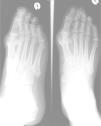
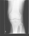
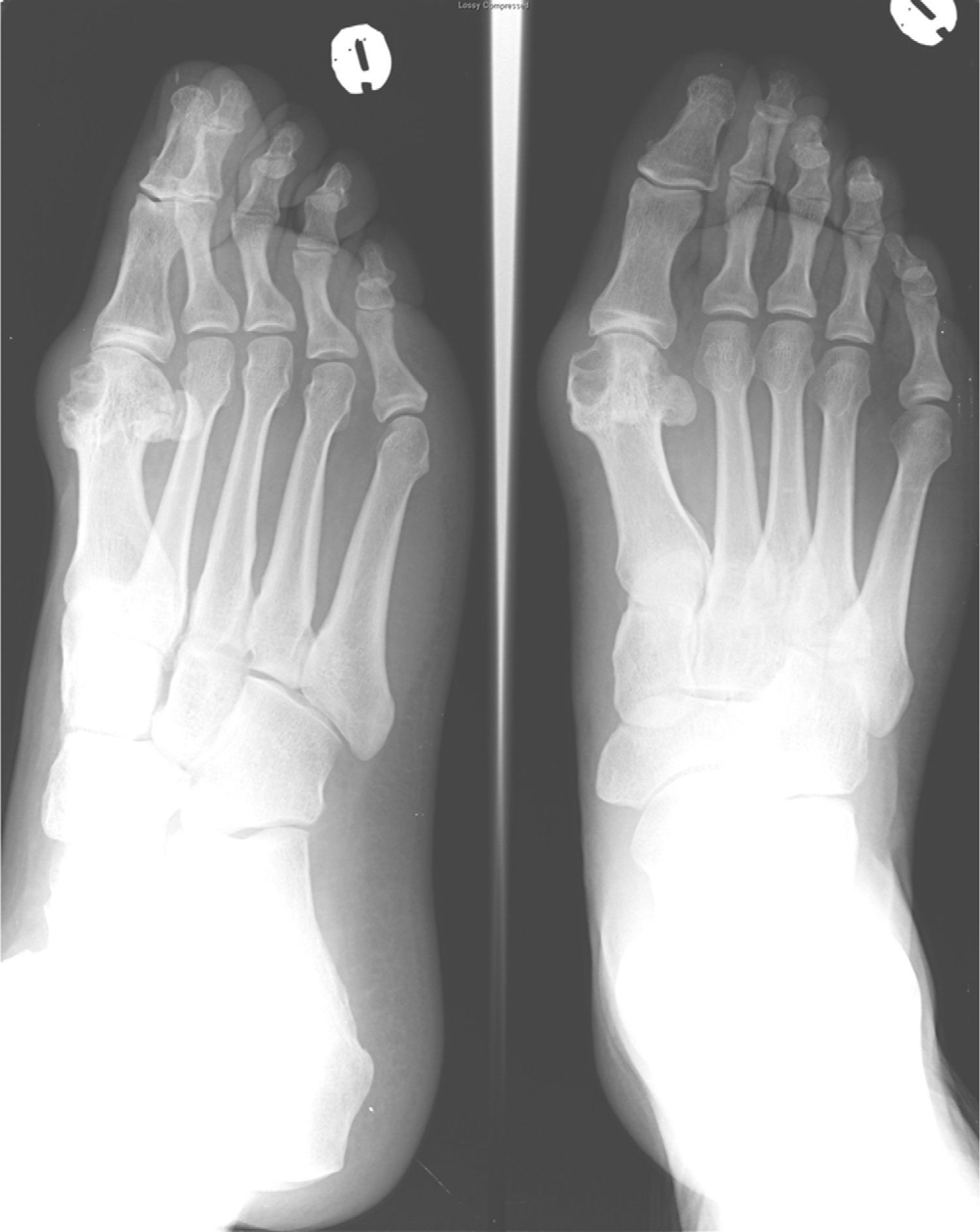
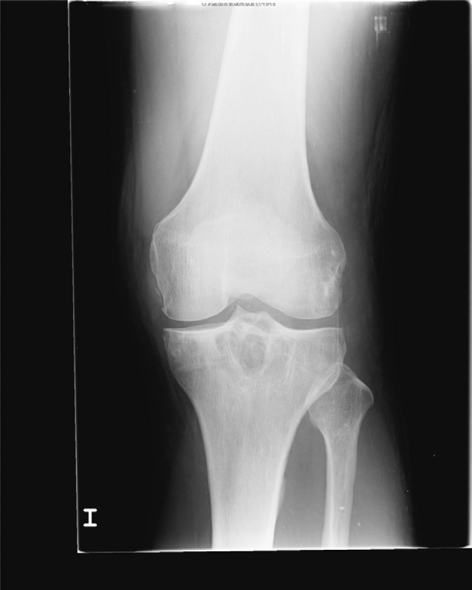
Laboratory tests showed uricemia of 9.4mg/dl and GGT 166U/dl. The remaining parameters (CBC, TSH, CPK, rheumatoid factor, HLA B27, anti-CCP antibodies, immunoglobulins, 24h urine uric acid) were normal. X-rays of the left foot were performed (Fig. 1), showing marginal erosions and sclerotic borders of the first metatarsal head, with increased adjacent soft tissue. A left knee radiograph (Fig. 2) showed lytic lesions with well-defined edges and incipient degenerative changes of the tibial epiphysis at the femorotibial level. We performed arthrocentesis of the left knee. Monosodium urate crystals were found in synovial fluid (SF). SF culture was negative.

Fig. 1.

Anteroposterior and oblique X-ray of left foot: well demarcated marginal erosions are observed on the first metatarsal head, with increased adjacent soft tissue. The joint space and the density of the rest of the subchondral bone is preserved.

(0.15MB).
Fig. 2.

Anteroposterior X-ray of the left knee: well demarcated lytic lesions are objectified in tibial epiphysis with incipient femorotibial degenerative changes.

(0.07MB).
Diagnosis and Evolution

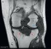
Given the radiological lesions of the left knee, we completed the study with a left knee MRI (Fig. 3) which showed synovial proliferation which revealed diffuse, hypointense lesions on T1, with bone erosions in the posterolateral margin of the femoral condyle. A large Baker's cyst with areas of high and low signal on T2 were seen, as well as lytic, hypointense lesion on T1, T2 with a heterogeneous signal level on the proximal tibial epiphysis and a maximum diameter of 5cm that after administration of gadolinium showed enhancement, something characteristic of tophaceous deposits.

Fig. 3.

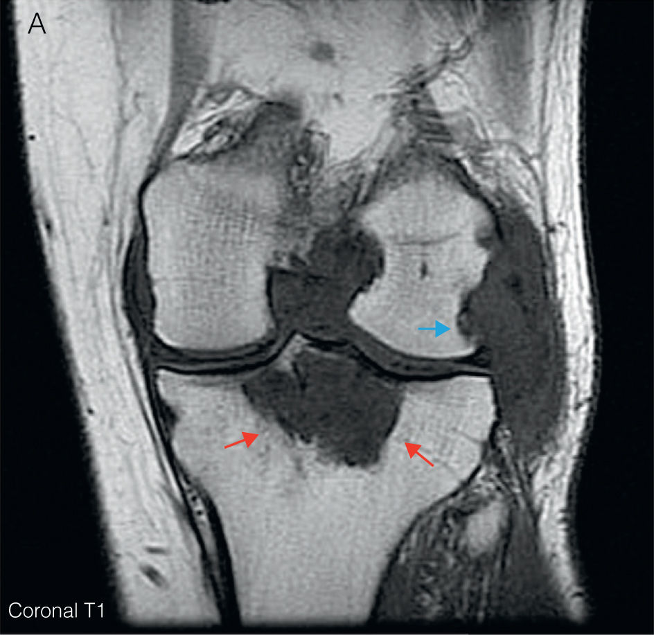
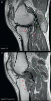
(A) Left knee coronal T1MRI. It shows a diffuse hypointense synovial proliferation with bone erosion of the posterolateral margin of the femoral condyle (blue arrow) and intercondylar notch. A hypointense lytic lesion is seen in the proximal tibial epiphysis with a maximum diameter of 5cm (red arrows). (B) Sagittal T2. There is an increased signal heterogeneity at the level of the tibial epiphyseal lytic lesion (red arrows), along with a large Baker's cyst located behind the medial gastrocnemius muscle (green arrows), with areas of high and low signal for synovial hypertrophy. (C) Sagittal T1-Gd. After administration of gadolinium, enhancement shows the level of peripheral tibial lytic lesion (red arrows) and Baker's cyst (green arrows). (For interpretation of the references to color in this figure legend, the reader is referred to the web version of the article.)

(0.12MB).

Based on the above, the patient was diagnosed with chronic gouty arthritis with intraosseous and intraarticular tophi. Dietary measures were initiated and treatment begun with allopurinol (300mg/d) and colchicine 1mg daily. Due to the persistence of clinical uricemia of 6.2mg/dl, benzbromarone was added (up to 100mg doses per day), until the patient was asymptomatic. Current levels of uric acid are 3.6mg/dl and there was a slight decrease of the tibial intraosseous tophus size.

Discussion

It is estimated that the prevalence of gouty arthritis in the general population is 1.4%,1 and represents approximately 5% of monoarthritis seen in consultation. The tophus is a nodular mass composed of sodium urate salts and surrounded by a chronic inflammatory reaction with abundant vascularization. It is a late complication of hyperuricemia maintained and present in only a minority of patients. They usually form in juxta-articular regions, although virtually any tissue2 can present them. It is not uncommon for intraosseous tophi to form in advanced crystal arthropathy, reaching 13.3% in patients in some series.3 A definitive diagnosis requires histopathological confirmation, but imaging tests may be useful. Plain radiographs can show juxta-articular and intraosseous calcifications and erosions of the articular surface, creating problems of differential diagnosis with avascular4 necrosis and osteochondroma. The presence of bone erosions is a marker of intrabone5 tophi. These can be seen as a soft tissue or intraosseous mass. The joint space is generally respected and periosteal new bone formation and subchondral collapse can appear. Is a useful tool to determine the severity of involvement, with standardized methods and a Sharp/Van der Heijde6 score available for indexing. Ultrasound is a useful tool for its low cost, no radiation, and speed of implementation, also used to guide invasive techniques. It has a high sensitivity for detecting monosodium urate deposits, even in patients with asymptomatic hyperuricemia. It is able to appreciate bone erosions earlier than plain X-rays. On the other hand, it is the only standardized technique for measurement of tophi and can demonstrate subclinical inflammatory activity by capturing Doppler hypervascular areas around the monosodium urate deposits. The typical findings in gout with this technique are: a double contour sign, aggregated punctate echogenic tissue, “snow storm” synovial fluid appearance with hyperechogenic areas, and soft tissue edema, in addition to the aforementioned bone erosions and hypervascularity.6–8 Computed tomography (CT) is useful in the recognition of monosodium urate deposits. These deposits have an attenuation of approximately 160 Hounsfield units (HU), while calcium crystals exhibit an attenuation of 450HU, even bone tissue and calcified tophi.8 It provides more specific images than ultrasound or MRI, as well as aid in the diagnosis of complications, and intraosseous tophi and bone erosions, and can guide joint aspiration.6 Magnetic resonance imaging (MRI) is a tool that, while non-specific and with certain limitations, such as suboptimal definition for intraosseous abnormalities, a high cost, and poor availability, is the method of choice for evaluating soft tissue, ligaments, muscles and tissues as well as juxta-articular joint space, and effusions or synovial proliferation, cartilage abnormalities, due to a spatial resolution far superior to other imaging tests. In particular, it has great utility in determining the etiology of functional limitations of unknown causes in the knee joint in patients with gout.2,6 Importantly, the synovial membrane is too thin to be assessed with this imaging method, so that it can only be seen when a pathological process enlarges it. The differential diagnosis of synovial thickening raises the possibility of longstanding rheumatoid arthritis, pyrophosphate arthropathy, villonodular synovitis, hemophilic arthropathy, amyloid arthropathy, synovial chondromatosis, and chronic granulomatous processes such as arthritis or fungal infection or tuberculosis.2,6,9 Gout is often found presenting a synovial effusion and synovial pannus. The tophi are seen as an amorphous lesion, eccentric and asymmetric, with variable intensity depending on their composition: protein, fibrous crystals, hemosiderin, and calcium, although the latter are rare and found in very old lesions. It is common to observe one of these morphological patterns: amorphous mass, linear crystalline deposits, or cystic lesions in the juxta-articular bursa. Intraosseous tophi are hypointense or of intermediate intensity on T1, heterogeneous-looking from hyper-to hypointense on T2, depending on the amount of calcium in the tophi, with more calcium, less intensity. It usually enhances after contrast administration, which translates hypervascularization and is a predictor of bone erosion. It also often is accompanied by juxta-articular findings such as erosions, edema of soft tissues, bone marrow edema, or extraarticular2 tophi.3,6,9–11 Isolated tophi may pose problems in differential diagnosis with tumors such as fibroblastic tumors and benign xantofibroma, but it is rare to find surrounding edema in these.10

In short, chronic tophaceous gout presents a constellation of clinical and radiological findings which are important to know for a correct differential diagnosis and further treatment. Although MRI is not a specific test for the diagnosis of gout, it does allow early detection of intraosseous and intraarticular tophi and bone erosions in patients with gout, helping us to recognize this entity and avoid a bone biopsy in selected patients. Thus, MRI can play a special role in the identification of clinical complications and monitoring response to treatment.

Ethical disclosures

Protection of human and animal subjects. The authors declare that no experiments were performed on humans or animals for this investigation.

Confidentiality of Data. The authors declare that they have followed the protocols of their work centre on the publication of patient data and that all the patients included in the study have received sufficient information and have given their informed consent in writing to participate in that study.

Right to privacy and informed consent. The authors have obtained the informed consent of the patients and /or subjects mentioned in the article. The author for correspondence is in possession of this document.

References
[1]
T.G. Rider, K.M. Jordan.
The modern management of gout.
Rheumatology, 49 (2010), pp. 5-14
[2]
J. Llauger, J. Palmer, N. Rosón, S. Bagué, A. Camins, R. Cremades.
Nonseptic monoarthritis: imaging features with clinical and histopathologic correlation.
[3]
K.H. Ko, Y.C. Hsu, H.S. Lee, C.H. Lee, G.S. Huang.
Tophaceous gout of the knee: revisiting MRI patterns in 30 patients.
J Clin Rheumatol, 16 (2010), pp. 209-214
[4]
D. Resnick, T.W. Broderick.
Intraosseous calcifications in tophaceous gout.
Am J Roentgenol, 137 (1981), pp. 1157-1161
[5]
N. Dalbeth, B. Clark, K. Gregory, G. Gamble, T. Sheehan, A. Doyle, et al.
Mechanisms of bone erosion in gout: a quantitative analysis using plain radiography and computed tomography.
Ann Rheum Dis, 68 (2009), pp. 1290-1295
[6]
F. Perez-Ruiz, N. Dalbeth, A. Urresola, E. De Miguel, N. Schlesinger.
Imaging of gout: findings and utility.
Arthritis Res Ther, 11 (2009), pp. 232
[7]
R.G. Thiele.
Role of ultrasound and other advanced imaging in the diagnosis and management of gout.
Curr Rheumatol Rep, 13 (2011), pp. 146-153
[8]
J.C. Gerster, M. Landry, L. Dufresne, J.Y. Meuwly.
Imaging of tophaceous gout: computed tomography provides specific images compared with magnetic resonance imaging and ultrasonography.
Ann Rheum Dis, 61 (2002), pp. 52-54
[9]
J.A. Narváez, J. Narváez, R. Ortega, E. De Lama, Y. Roca, N. Vidal.
Hypointense synovial lesions on T2-weighted images: differential diagnosis with pathologic correlation.
AJR Am J Roentgenol, 181 (2003), pp. 761-769
[10]
J.S. Yu, C. Chung, M. Recht, T. Dailiana, R. Jurdi.
MR imaging of tophaceous gout.
Am J Roentgenol, 168 (1997), pp. 523-527
[11]
F. Paparo, G. Zampogna, E. Fabbro, M. Parodi, R. Andracco, G. Ferrero, et al.
Imaging of tophi with an extremity-dedicated MRI system.
Clin Exp Rheumatol, 29 (2011), pp. 519-526

Please cite this article as: Rodríguez Leal CM, et al. Tofo tibial intraóseo en gota crónica. Reumatol Clin. 2012. doi:10.1016/j.reuma.2011.10.010.

Copyright © 2011. Elsevier España, S.L.. All rights reserved
Descargar PDF
Idiomas
Reumatología Clínica
Opciones de artículo
Herramientas
es en

¿Es usted profesional sanitario apto para prescribir o dispensar medicamentos?

Are you a health professional able to prescribe or dispense drugs?