A 37-year-old woman with a fibrous polyostotic dysplasia (FPD) of the left femur, tibia and foot was diagnosed at 11 years of age. At the onset she presented mechanical pain of the left hip and later a stress fracture of the femur for which she was treated with surgery, pamidronate and zolendronate. Pathology confirmed the diagnosis. Phosphocalcic metabolism was normal.
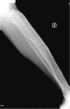
X-rays (Fig. 1) showed a left femur with a «sheperd's staff » deformity, a thin bone cortex and expansive radiolucent lesions. The left tibia (Fig. 2) was curved and had a thin cortex. Feet (Fig. 3) were thickened at the first right metatarsal and phalange with radiolucent and sclerotic areas. A computed tomography of the tibia (Fig. 4) observed a ground glass matrix, with heterogeneous intramarrow images.
FPD is a rare anomaly of skeletal development. A mutation in the GNAS1 gene has been detected,1 producing alterations in osteoplastic maturation and abnormal fibrous tissue deposit.2 There are two variants: monostotic and polyostotic.3 Lesions are localized on the epiphysis, metaphysis or diaphysis.
The monostotic variant is more prevalent, diagnosed during the patient's youth and less symptomatic. It affects the ribs, femur, tibia, jawbone and humerus.4
The polyostotic form is observed in 30% of cases. It is usually diagnosed during the patients’ infancy. It affects the cranium, face, pelvis, spine and shoulder. It is associated to the McCune-Albright syndrome in 2% of cases (FPD, skin pigmentation and early puberty).2 It leads to dysmetria, gait abnormalities, mechanical pain and stress fractures.5 FPD diagnosis is radiological, rarely requiring a bone biopsy. The prognosis depends on the extension and degree of bone affection, age at onset and extraskeletal manifestations.2 The malignity rate is rare.6
In case of pain, deformity or fracture, treatment with biphosphonate is recommended.7 Curetage or bone implants might be necessary.
Didactic MessageIn young patients with bone deformity, PFD must be considered as a diagnosis. Simple X-rays might be enough for diagnosis.
Ethical ResponsibilitiesProtection of People and AnimalsThe authors state that no experiments were performed on people or animals for this study.
Confidentiality of DataThe authors state that the protocols of their center regarding the publication of patient data have been followed.
Right to Privacy and Informed ConsentThe authors have obtained informed consent from the patients and/or subjects referred to in the article. This document is in the possession of the corresponding author.
Please cite this article as: Meneses CF, Egües A, Uriarte M, Belzunegui J. Displasia fibrosa poliostótica: presentación de un caso. Reumatol Clin. 2014;10:413–415.


