Non-traumatic acute shoulder pain occupies an important place within emergency hospital visits, accounting for about 7% of all visits due to locomotor system problems.1 Ultrasound scan is an extremely useful tool in the study of this complaint, complementing the preparation of a correct clinical history and detailed physical examination, permitting easily accessed confirmatory etiological identification with a precision comparable to that of magnetic resonance imaging.2
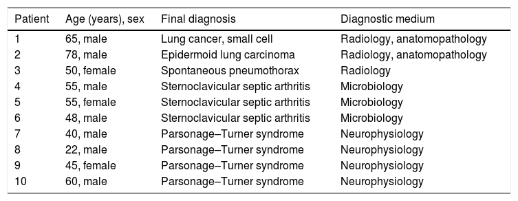
In general terms the most frequent cause of chronic shoulder pain is tendon disease (or tendinopathy). This definition covers all disease that alters fibrous tendon architecture.2,3 While partial and complete tendon tears are often found in elderly patients, with or without fatty muscular degeneration, tendinosis tends to be found more often in younger patients (defined as the presence of changes in echogenicity in the fibrous structure or thickening of part or all of the tendon), or partial tears that break the continuity of the fibres.3–5 Structural changes, fundamentally tendinous ones, can be detected in patients with acute or chronic omalgia, while in acute cases it is far more common to observe bursitis or bleeding in the biceps sheath.6 Nevertheless, it is rare to encounter cases of acute shoulder pain in which the ultrasound scan of the shoulder is normal. In our recent experience we have identified 10 cases of patients with acute omalgia and normal shoulder ultrasound scan results whose final diagnoses were especially alarming. We believe it is relevant to report these cases due to their clinical importance (Table 1).
Summarised description of cases of shoulder pain with a normal ultrasound scan.
| Patient | Age (years), sex | Final diagnosis | Diagnostic medium |
|---|---|---|---|
| 1 | 65, male | Lung cancer, small cell | Radiology, anatomopathology |
| 2 | 78, male | Epidermoid lung carcinoma | Radiology, anatomopathology |
| 3 | 50, female | Spontaneous pneumothorax | Radiology |
| 4 | 55, male | Sternoclavicular septic arthritis | Microbiology |
| 5 | 55, female | Sternoclavicular septic arthritis | Microbiology |
| 6 | 48, male | Sternoclavicular septic arthritis | Microbiology |
| 7 | 40, male | Parsonage–Turner syndrome | Neurophysiology |
| 8 | 22, male | Parsonage–Turner syndrome | Neurophysiology |
| 9 | 45, female | Parsonage–Turner syndrome | Neurophysiology |
| 10 | 60, male | Parsonage–Turner syndrome | Neurophysiology |
Two patients, both males aged 65 and 78 years old, had visited due to shoulder pain with mechanical characteristics. Both of them had been examined radiologically and they had received conservative treatment, with rest and first level analgesics. The 78-year-old male had asymmetry of the right supraclavicular triangle, corresponding to the painful shoulder (Fig. 1A and B). After a normal ultrasound scan of the shoulder, both patients were found to have primary tumours of the lung.
Patients with acute shoulder pain and normal ultrasound scan results. (A) Appearance of the supraclavicular regions of the male patient aged 78 years old, clearly showing the asymmetry of the right supraclavicular triangle in comparison with the left one. (B) Thoracic X-ray image of the same patient, showing a pulmonary mass subsequently identified as epidermoid carcinoma. (C) Posteroanterior X-ray image of a 50-year-old woman with acute shoulder pain, showing a complete pneumothorax of the right hemithorax without mediastinal deviation.
A 50-year-old Asiatic woman visited due to right shoulder pain. The physical examination and ultrasound scan of the shoulder were both normal. Given the lack of correspondence between the intensity of the symptoms and the normality of the ultrasound scan an X-ray was taken which showed right pneumothorax (Fig. 1C).
Two patients, a 55-year-old man and a woman of the same age and both immunocompetent, originally visited due to recent mechanical omalgia. They were eventually diagnosed as cases of sternoclavicular septic arthritis. Both cases were exhaustively described beforehand.7 A third patient, a 48-year-old male, with a history of infection by human immunodeficiency virus and in antiretroviral treatment had sternoclavicular septic arthritis 2 weeks after having commenced with ipsilateral omalgia that a week afterwards led to study of the shoulder by ultrasound scan that was reported as normal.
Three patients, 2 men aged 40 and 22 years old, and a woman aged 45 years old, whose cases were described beforehand by our group,8 visited due to acute shoulder pain with major functional repercussion. In all 3 cases the ultrasound scan report was normal. After neurophysiological studies they were diagnosed with Parsonage–Turner syndrome (PTS). Recently, a 60-year-old male patient who had recently received an anti-tetanus vaccination with immunoglobulin, had the same symptoms with normal results of ultrasound scan of the shoulder. The electrophysiological study was compatible with PTS. In our accumulated experience from 2011 to 2017, the volume of normal ultrasound scan reports corresponding to patients who visited the emergency department with acute shoulder pain (arbitrarily considered to have evolved during less than 3 weeks, due to the characteristics of the demand for care in our hospital) represents about 7% of all ultrasound explorations. The 10 cases we describe in this letter represent approximately one third of our normal number of normal reports. We believe that it is important to underline that in cases of acute omalgia, after a detailed clinical history and meticulous physical examination, that a normal ultrasound scan report should be followed by a broader diagnostic study, given that some differential diagnoses of shoulder pain with normal ultrasound scan results require prompt therapeutic intervention.
Please cite this article as: Guillen Astete CA, Garcia Garcia V, Luque Alarcon M. Importancia de la ecografía normal en pacientes con dolor agudo de hombro de origen no traumático. Reumatol Clin. 2020;16:509–510.


